

Sox2 and Pou2f1 interact to control lens and olfactory placode development
- PMID: 17140559
- PMCID: PMC3276313
- DOI: 10.1016/j.ydbio.2006.10.047
Sox2 and Pou2f1 interact to control lens and olfactory placode development
Abstract
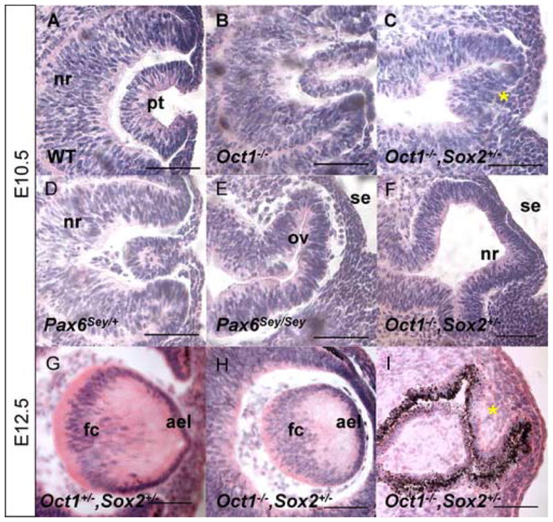
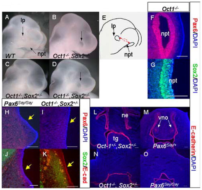
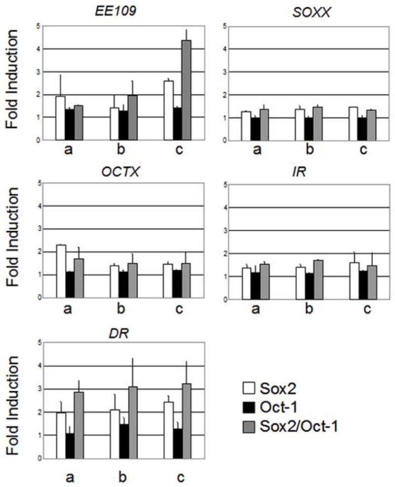
Sox2, which encodes an SRY-like HMG box transcription factor, is critical for vertebrate development. Sox2 mediates its transcriptional effects through the formation of complexes with specific co-factors, many of which are unknown. In this report, we identify Oct-1, encoded by the Pou2f1 gene, as a co-factor for Sox2 in the context of mouse lens and nasal placode induction. Oct-1, Sox2, and Pax6 are co-expressed during lens and nasal placode induction and during subsequent developmental stages. Genetic combination of Sox2 and Pou2f1 mutant alleles results in impaired induction of the lens placode, an ocular phenotype that includes anophthalmia, and a complete failure of nasal placode induction. These ocular and nasal phenotypes closely resemble those observed in Pax6 null embryos. Moreover, we identify DNA-binding sites that support the cooperative formation of a complex between Sox2 and Oct-1 and mediate Sox2/Oct-1-dependent transactivation of the Pax6 lens ectoderm enhancer in vitro. We demonstrate that the same Sox- and Octamer-binding sites are essential for Pax6 enhancer activity in the lens placode and its derivatives in transgenic mouse embryos. Collectively, these results indicate that Pou2f1, Sox2 and Pax6 are interdependent components of a molecular pathway utilized in both lens and nasal placode induction.
Figures


Similar articles
-
Stage-dependent modes of Pax6-Sox2 epistasis regulate lens development and eye morphogenesis.Development. 2009 Sep;136(17):2977-85. doi: 10.1242/dev.037341. Development. 2009. PMID: 19666824 Free PMC article.
-
Pax6 and SOX2 form a co-DNA-binding partner complex that regulates initiation of lens development.Genes Dev. 2001 May 15;15(10):1272-86. doi: 10.1101/gad.887101. Genes Dev. 2001. PMID: 11358870 Free PMC article.
-
PAX6 and SOX2-dependent regulation of the Sox2 enhancer N-3 involved in embryonic visual system development.Genes Cells. 2007 Sep;12(9):1049-61. doi: 10.1111/j.1365-2443.2007.01114.x. Genes Cells. 2007. PMID: 17825048
-
Interplay of Pax6 and SOX2 in lens development as a paradigm of genetic switch mechanisms for cell differentiation.Int J Dev Biol. 2004;48(8-9):819-27. doi: 10.1387/ijdb.041868hk. Int J Dev Biol. 2004. PMID: 15558474 Review.
-
Regulation of gene expression by Pax6 in ocular cells: a case of tissue-preferred expression of crystallins in lens.Int J Dev Biol. 2004;48(8-9):829-44. doi: 10.1387/ijdb.041866ac. Int J Dev Biol. 2004. PMID: 15558475 Free PMC article. Review.
Cited by
-
SP8 regulates signaling centers during craniofacial development.Dev Biol. 2013 Sep 15;381(2):312-23. doi: 10.1016/j.ydbio.2013.07.007. Epub 2013 Jul 18. Dev Biol. 2013. PMID: 23872235 Free PMC article.
-
Identification of in vivo DNA-binding mechanisms of Pax6 and reconstruction of Pax6-dependent gene regulatory networks during forebrain and lens development.Nucleic Acids Res. 2015 Aug 18;43(14):6827-46. doi: 10.1093/nar/gkv589. Epub 2015 Jul 2. Nucleic Acids Res. 2015. PMID: 26138486 Free PMC article.
-
Parent-of-origin effects in SOX2 anophthalmia syndrome.Mol Vis. 2011;17:3097-106. Epub 2011 Nov 24. Mol Vis. 2011. PMID: 22171155 Free PMC article.
-
Mechanisms controlling diversification of olfactory sensory neuron classes.Cell Mol Life Sci. 2017 Sep;74(18):3263-3274. doi: 10.1007/s00018-017-2512-2. Epub 2017 Mar 29. Cell Mol Life Sci. 2017. PMID: 28357469 Free PMC article. Review.
-
Structural basis for nuclear import selectivity of pioneer transcription factor SOX2.Nat Commun. 2021 Jan 4;12(1):28. doi: 10.1038/s41467-020-20194-0. Nat Commun. 2021. PMID: 33397924 Free PMC article.
References
-
- Ahrens K, Schlosser G. Tissues and signals involved in the induction of placodal Six1 expression in Xenopus laevis. Dev Biol. 2005;288:40–59. - PubMed
-
- Aota S, Nakajima N, Sakamoto R, Wantabe S, Ibarake N, Okazaki K. Pax6 autoregulation mediated by direct interaction of Pax6 protein with the head surface ectoderm-specific enhancer of the mouse Pax6 gene. Dev Biol. 2003;257:1–13. - PubMed
-
- Ashery-Padan R, Gruss P. Pax6 lights-up the way for eye development. Curr Opin Cell Biol. 2001;13:706–714. - PubMed
Publication types
MeSH terms
Substances
Grants and funding
LinkOut - more resources
Full Text Sources
Molecular Biology Databases
Miscellaneous
