

doi: 10.1016/j.semcdb.2006.10.007.
Epub 2006 Oct 26.
Sef and Sprouty expression in the developing ocular lens: implications for regulating lens cell proliferation and differentiation
Affiliations
- PMID: 17141539
- PMCID: PMC1847394
- DOI: 10.1016/j.semcdb.2006.10.007
Item in Clipboard
Sef and Sprouty expression in the developing ocular lens: implications for regulating lens cell proliferation and differentiation
Semin Cell Dev Biol.
2006 Dec.
Abstract
In many developmental systems, growth factor signalling must be temporally and spatially regulated, and this is commonly achieved by growth factor antagonists. Here, we describe the expression patterns of newly identified growth factor inhibitors, Sprouty and Sef, in the developing ocular lens. Sprouty and Sef are both expressed in the lens throughout embryogenesis, and become restricted to the lens epithelium, indicating that lens cell proliferation and fibre differentiation may be tightly regulated by such antagonists. Future studies will be aimed at understanding how these negative regulatory molecules modulate growth factor-induced signalling pathways and cellular processes in the lens.
Figures
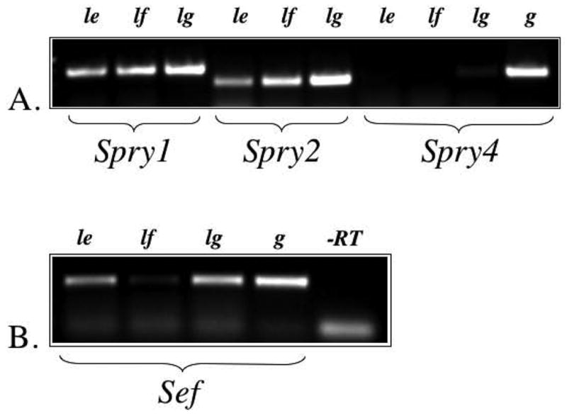
Expression of Spry1, Spry2, Spry4 and Sef using RT-PCR. A. Spry1 and Spry2, but not Spry4, were detected in the rat lens epithelium (le) and lens fibre cells (lf). Lung tissue (lg), and genomic DNA (g) were used as positive controls. B. Sef was detected in the lens epithelium, lens fibre cells (weakly) and lung. Genomic DNA was used as a positive control, and reverse transcriptase (-RT) was omitted to control for DNA contamination of RNA samples.
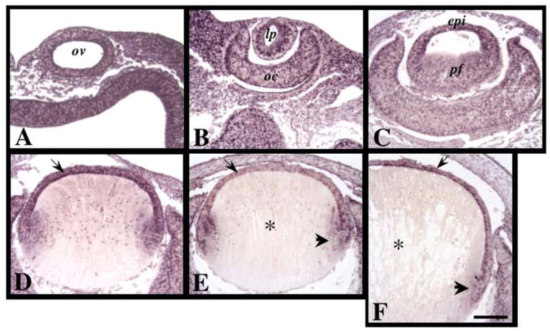
Expression of Spry1 mRNA during murine embryonic lens morphogenesis. Sections of mouse eyes at embryonic stages E9.5 (A), E10.5 (B), E12.5 (C), E13.5 (D), E14.5 (E) and E15.5 (F) were labelled for Spry1 transcripts using in situ hybridisation. Spry1 was ubiquitously expressed early in development (A–C). By E13.5, strongest expression for Spry1 was detected in the anterior monolayer of lens cells (D, arrow). At E14.5 and E15.5, Spry1 was expressed in the epithelial cells (E, F, arrow) and early secondary fibre cells (E, F, arrowhead), but not in the maturing primary fibre cells (E, F, asterisks). Abbreviations: epi, lens epithelium; lp, lens pit; oc, optic cup; ov, optic vesicle; pf, primary fibre cells. Scale bar: 100 μm.
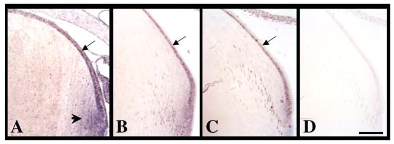
Expression of Spry1 mRNA during postnatal murine lens growth. Sections of eyes at postnatal stages P0 (A), P14 (B) and P21 (C, D) were labelled for Spry1 transcripts using in situ hybridisation. (A) At P0, Spry1 was strongly expressed in lens epithelial cells (arrow) and early secondary fibre cells (arrowhead). At P14 and P21, Spry1 was primarily expressed in the lens epithelial cells (B, C, arrows). (D) Control sections using a sense-labelled riboprobe showed no signal. Scale bar: 100 μm.
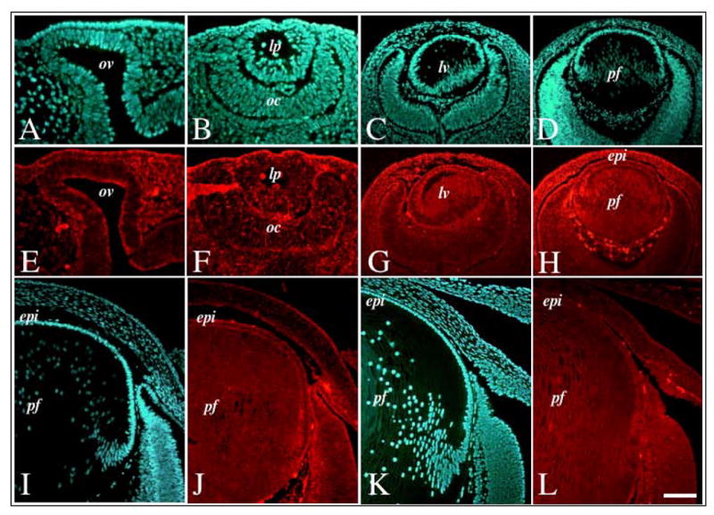
Expression of Spry2 mRNA during murine embryonic lens morphogenesis. Sections of mouse eyes at embryonic stages E9.5 (A, E), E10.5 (B, F), E12.5 (C, G), E13.5 (D, H), E15.5 (I, J) and E18.5 (K, L) were labelled for Spry2 transcripts using fluorescence in situ hybridisation (E–H, J, L) and counterstained with Hoechst dye (A–D, I, K). Spry2 was ubiquitously expressed throughout lens morphogenesis, with strong expression in both the lens epithelium and primary fibre cells. Abbreviations: epi, lens epithelium; lp, lens pit; lv, lens vesicle ; oc, optic cup; ov, optic vesicle; pf, primary fibres. Scale bar: 50 μm (A, B, E, F); 100 μm (C, D, G, H); 80 μm (I–L).
Expression of Spry2 mRNA during postnatal murine lens growth. Sections of eyes at postnatal stages P3 (A, D, G, J), P15 (B, E, H, K) and P21 (C, F, I, L) were labelled for Spry2 transcripts using either fluorescent in situ hybridisation (A–C) or alkaline-phosphatase labelled riboprobes (D–L). At P3, Spry2 was expressed throughout the lens, in both epithelial cells (A, D, G, arrows) and fibre cells (A, D, G, asterisk). At P15 and P21, Spry2 expression in the lens epithelium (B, C, E, F, H, I, arrow) was retained, but progressively lost in the maturing lens fibre cells (B, C, E, F, H, I, asterisk). Control sections using a sense-labelled riboprobe showed no signal (J–L). Scale bar: 100 μm.
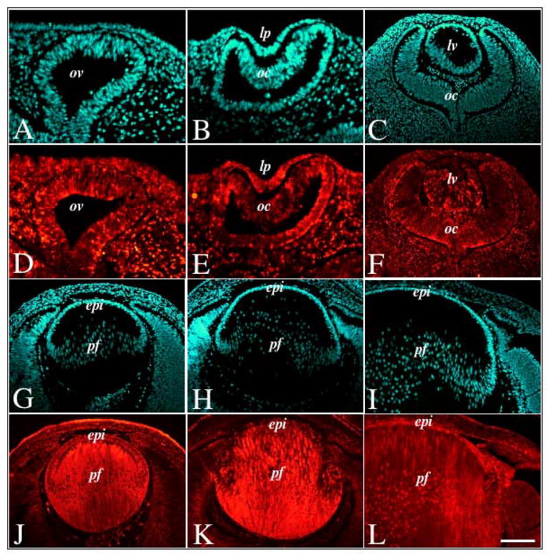
Immunofluorescent labelling of Spry2 during murine embryonic lens morphogenesis. Sections of mouse eyes at embryonic stages E11.5 (A, B, D, E), E12.5 (C, F), E13.5 (G, J), E15.5 (H, K) and E18.5 (I, L) were immunolabelled for Spry2 protein (D–F, J–L) and counterstained with Hoechst dye (A–C, G–I). Spry2 protein was ubiquitously expressed throughout lens morphogenesis, with strong expression in both the lens epithelium and primary fibre cells. Abbreviations: epi, lens epithelium; lp, lens pit; lv, lens vesicle; oc, optic cup; ov, optic vesicle; pf, primary fibres. Scale bar: 100 μm.
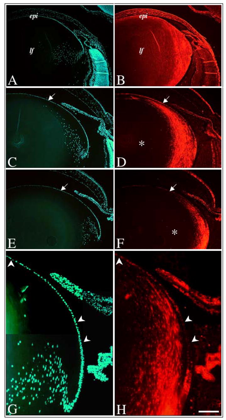
Immunofluorescent labelling of Spry2 during postnatal murine lens growth. Sections of mouse eyes at postnatal stages P3 (A, B), P10 (C, D), and P21 (E, F–H), were immunolabelled for Spry2 protein (B, D, F, H) and counterstained with Hoechst dye (A, C, E, G). In neonates, Spry2 labelling was strong throughout the lens, in both epithelial and fibre cells (B). At P10 (D) and P21 (F), Spry2 labelling was strong in the lens epithelium (arrows), but progressively lost in the 26 maturing lens fibre cells (asterisk). At higher magnification (G, H), the heterogenous labelling for Spry2 protein is more prominent with many lens epithelial cells demonstrating weak to reduced labelling (arrowheads). Abbreviations: epi, lens epithelium; lf, lens fibres. Scale bar: A–F, 200 μm, G–H, 100 μm.
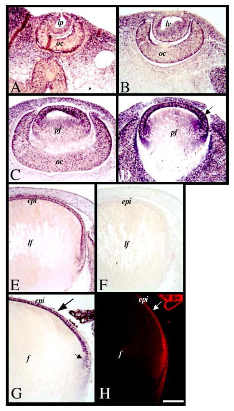
Expression of Sef mRNA during lens morphogenesis and growth. Sections of mouse eyes at embryonic stages E10.5 (A), E11.5 (B), E12.5 (C), E13.5 (D), E15.5 (E, F) and mouse postnatal stage P1 (G) and rat postnatal stage P21 (H) were labelled for Sef transcripts using alkaline phosphatase-labelled (A–G) or fluorescent (H) in situ hybridisation. Sef was ubiquitously expressed early in development (A–C). By E13.5 (D), strongest expression for Sef was detected in the differentiating anterior monolayer of lens cells (arrow). At E15.5, Sef was expressed in the epithelial cells, and not in the fibre cells. This labelling pattern persisted into postnatal development at birth (G) and weaning age (H). Control sections using a sense-labelled riboprobe showed no signal (F). Abbreviations: epi, lens epithelium; lp, lens pit; lv, lens vesicle; oc, optic cup; ov, optic vesicle; pf, primary fibre cells. Scale bar: 100 μm.
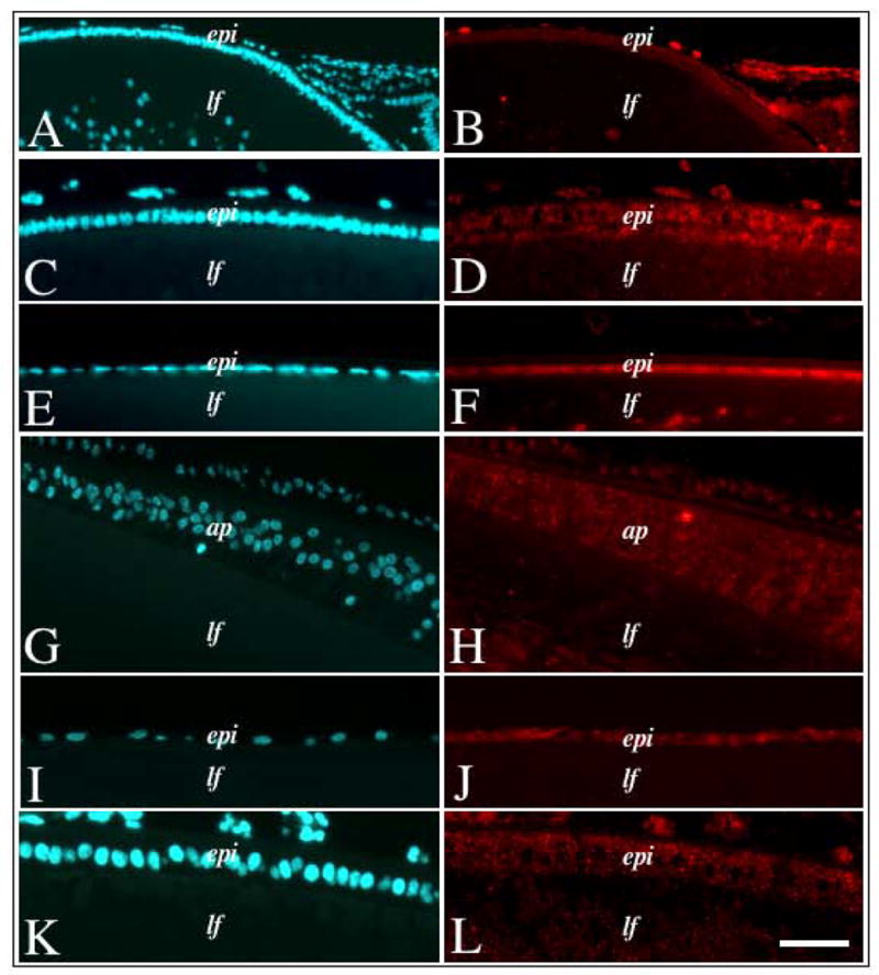
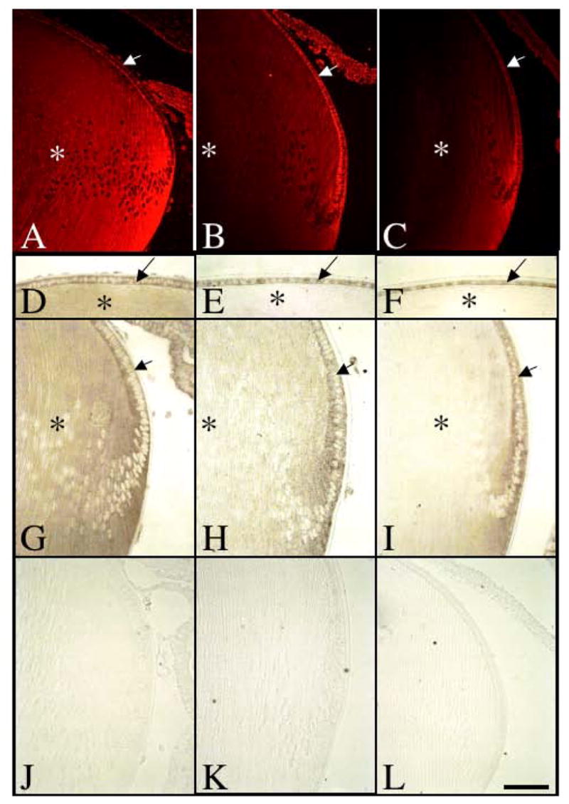
Immunofluorescent labelling of Sef in lenses of different species. Lens sections from neonatal mouse (A, B), neonatal rat (C, D), weanling rat (E, F), newborn chick (G, H), adult bovine (I, J) and foetal human (K, L) eyes, either immunolabelled for Sef protein (B, D, F, H, J, L) or counterstained with Hoechst dye (A, C, E, G, I, K). In all vertebrates examined, Sef protein labelling was strongest in the epithelial cells with little to no labelling in the fibre cells. Abbreviations: epi, lens epithelium; lf, lens fibres, ap, chick annular pad. Scale bar: 100 μm (A, B), 50 μm (C–L).
References
-
- McAvoy JW, Chamberlain C, de Iongh R, Hales A, Lovicu FJ. Lens Development. Eye. 1999;13:425–37. - PubMed
-
- Robinson ML, Lovicu FJ. The Lens: Historical and Comparative Perspectives. In: Lovicu FJ, Robinson ML, editors. Development of the Ocular Lens. New York: Cambridge University Press; 2004. pp. 3–26.
-
- Lovicu FJ, McAvoy JW. Growth Factor Regulation Of Lens Development. Dev Biol. 2005;280:1–14. - PubMed
-
- Schulz MW, Chamberlain CG, de Iongh RU, McAvoy JW. Acidic and basic FGF in ocular media and lens: implications for lens polarity and growth patterns. Development. 1993;118:117–26. - PubMed
-
- McAvoy JW, Chamberlain CG. Fibroblast growth factor (FGF) induces different responses in lens epithelial cells depending on its concentration. Development. 1989;107:221–28. - PubMed
Publication types
MeSH terms
Substances
Grants and funding
LinkOut - more resources
Full Text Sources
Molecular Biology Databases
