

Macrophage inflammatory protein 3alpha deficiency in atopic dermatitis skin and role in innate immune response to vaccinia virus
- PMID: 17141855
- PMCID: PMC2746067
- DOI: 10.1016/j.jaci.2006.10.005
Macrophage inflammatory protein 3alpha deficiency in atopic dermatitis skin and role in innate immune response to vaccinia virus
Erratum in
- J Allergy Clin Immunol. 2008 Nov;122(5):1007.. Kisich, Kevin [added]
Abstract
Background: Patients with atopic dermatitis (AD) are prone to disseminated viral skin infections and therefore are not vaccinated against smallpox because of potential complications. Macrophage inflammatory protein 3alpha (MIP-3alpha) is a C-C chemokine expressed by keratinocytes that exhibits antimicrobial activity against bacteria and fungi; however, its role in antiviral innate immunity is unknown.
Objective: Evaluate the level of MIP-3alpha in AD skin and its role in the innate immune response to vaccinia virus (VV).
Methods: Macrophage inflammatory protein 3alpha levels were evaluated using real-time RT-PCR, immunodot-blot, and immunohistochemistry. The antiviral activity of MIP-3alpha was determined using a standard viral plaque assay.
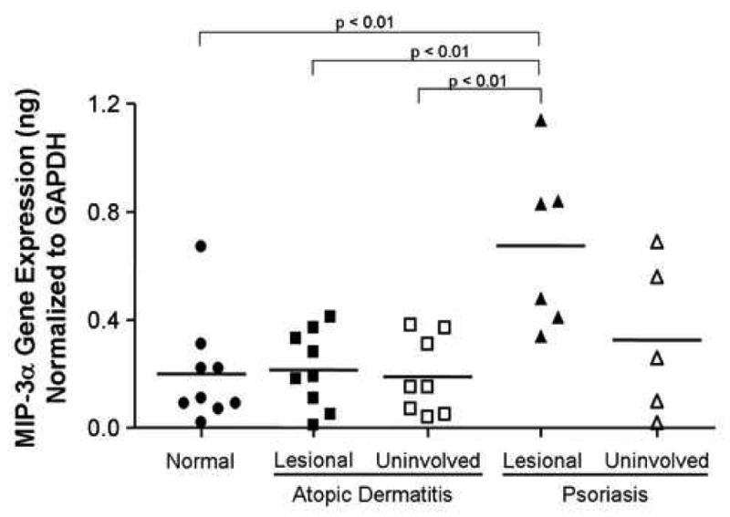
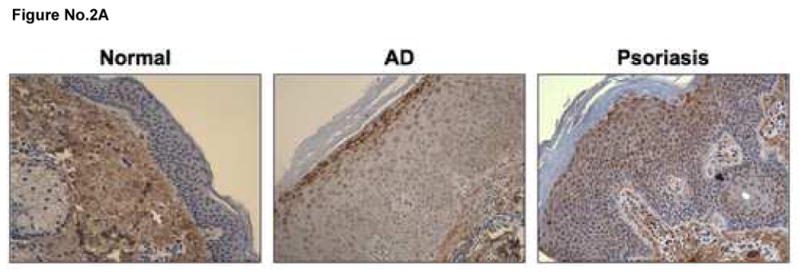
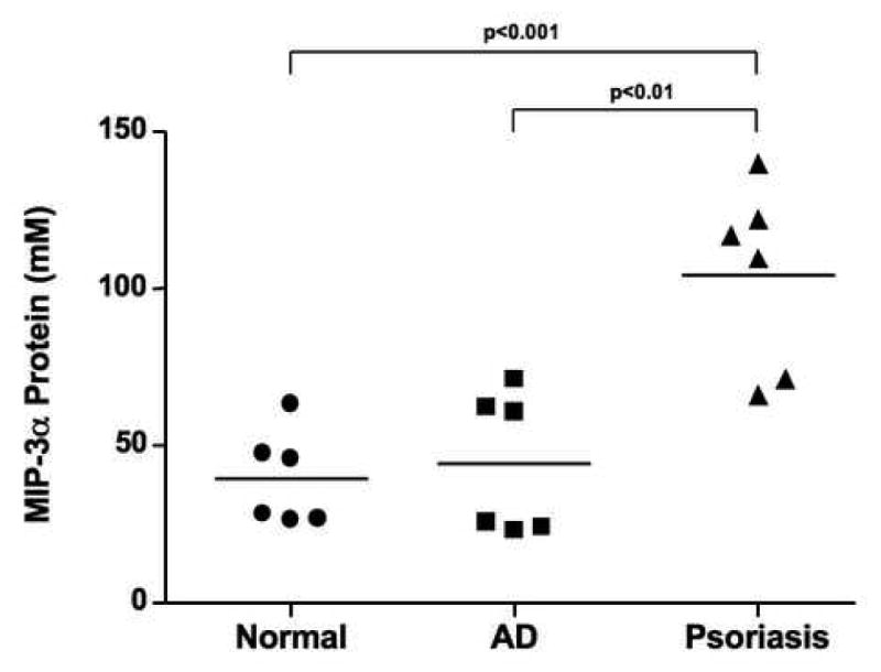
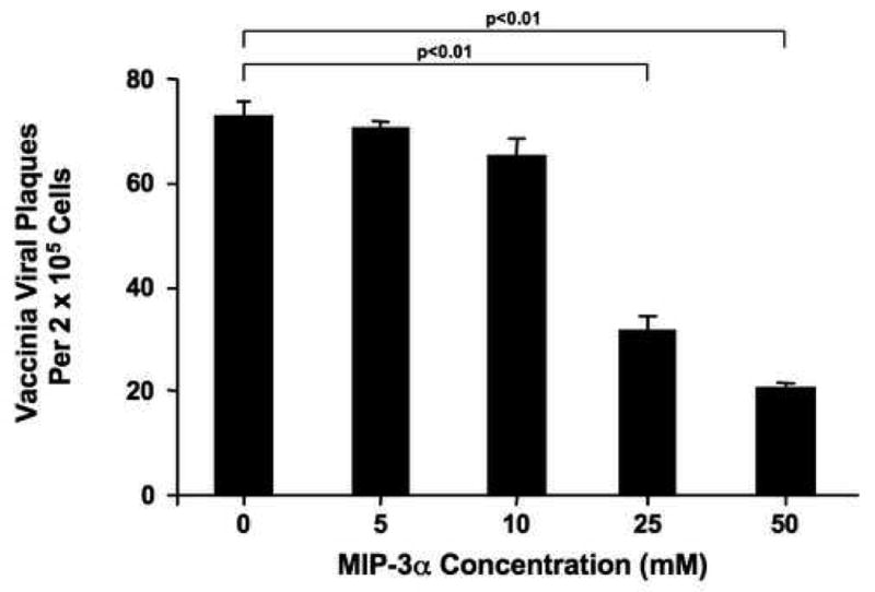
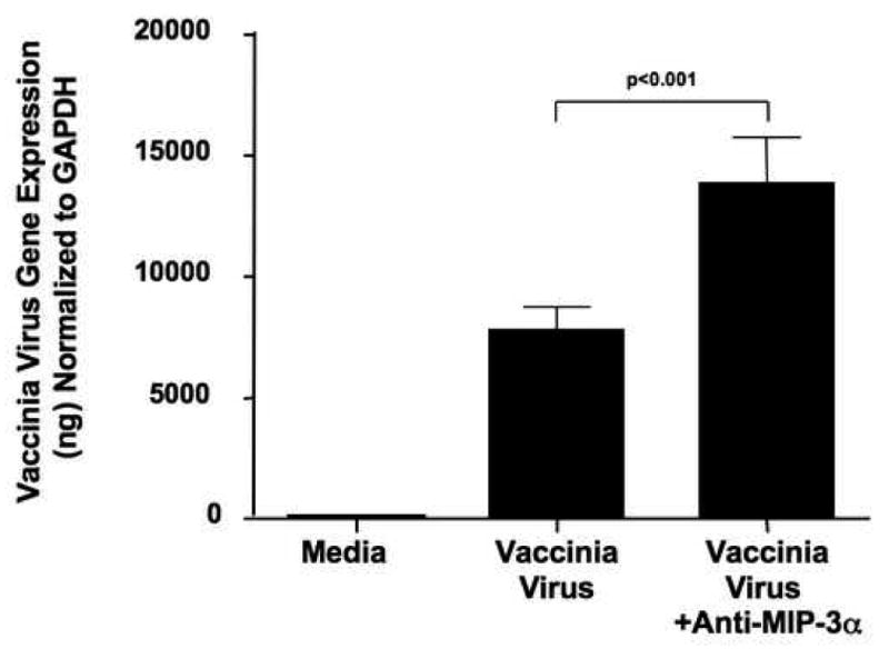
Results: Macrophage inflammatory protein 3alpha gene expression was significantly (P < .01) decreased in AD skin (0.21 +/- 0.05 ng MIP-3alpha/ng glyceraldehyde-3-phosphate dehydrogenase) compared with psoriasis skin (0.67 +/- 0.13). This was confirmed at the protein level using immunohistochemistry. We further demonstrate that T(H)2 cytokines downregulate MIP-3alpha expression. The importance of MIP-3alpha in the innate immune response against VV was established by first demonstrating that MIP-3alpha exhibits activity against VV. Second, VV replication was significantly increased (P < .01) in keratinocytes treated with an antibody to neutralize MIP-3alpha.
Conclusion: The current study demonstrates that MIP-3alpha exhibits antiviral activity against VV and demonstrates the importance of MIP-3alpha in the innate immune response against VV. In addition, AD skin is deficient in MIP-3alpha, in part because of the overexpression of T(H)2 cytokines in AD skin.
Clinical implications: MIP-3alpha deficiency in AD skin contributes to patients' increased propensity toward eczema vaccinatum. Increasing MIP-3alpha or neutralizing T(H)2 cytokines could prevent adverse reactions in patients with AD after smallpox vaccination.
Figures


Similar articles
-
Cytokine milieu of atopic dermatitis skin subverts the innate immune response to vaccinia virus.Immunity. 2006 Mar;24(3):341-8. doi: 10.1016/j.immuni.2006.02.006. Immunity. 2006. PMID: 16546102
-
Inducible expression of a CC chemokine liver- and activation-regulated chemokine (LARC)/macrophage inflammatory protein (MIP)-3 alpha/CCL20 by epidermal keratinocytes and its role in atopic dermatitis.Int Immunol. 2001 Jan;13(1):95-103. doi: 10.1093/intimm/13.1.95. Int Immunol. 2001. PMID: 11133838
-
Vaccinia virus-specific molecular signature in atopic dermatitis skin.J Allergy Clin Immunol. 2010 Jan;125(1):153-159.e28. doi: 10.1016/j.jaci.2009.10.024. J Allergy Clin Immunol. 2010. PMID: 20109744 Free PMC article.
-
Vaccinia virus pathogenicity in atopic dermatitis is caused by allergen-induced immune response that prevents the antiviral cellular and humoral immunity.Virus Genes. 2003 Dec;27(3):269-82. doi: 10.1023/a:1026399916888. Virus Genes. 2003. PMID: 14618088 Review.
-
Innate lymphoid cells and the skin.BMC Dermatol. 2014 Nov 26;14:18. doi: 10.1186/1471-5945-14-18. BMC Dermatol. 2014. PMID: 25427661 Free PMC article. Review.
Cited by
-
Antimicrobial activities of chemokines: not just a side-effect?Front Immunol. 2012 Jul 23;3:213. doi: 10.3389/fimmu.2012.00213. eCollection 2012. Front Immunol. 2012. PMID: 22837760 Free PMC article.
-
Modulation of Inflammatory Signaling Molecules in Bordetella pertussis Antigen-Challenged Human Monocytes in Presence of Adrenergic Agonists.Vaccines (Basel). 2022 Feb 17;10(2):321. doi: 10.3390/vaccines10020321. Vaccines (Basel). 2022. PMID: 35214778 Free PMC article.
-
Asthma and severity of 2009 novel H1N1 influenza: a population-based case-control study.J Asthma. 2013 Dec;50(10):1069-76. doi: 10.3109/02770903.2013.834505. Epub 2013 Sep 18. J Asthma. 2013. PMID: 23947393 Free PMC article.
-
Saliva-Derived Host Defense Peptides Histatin1 and LL-37 Increase Secretion of Antimicrobial Skin and Oral Mucosa Chemokine CCL20 in an IL-1α-Independent Manner.J Immunol Res. 2017;2017:3078194. doi: 10.1155/2017/3078194. Epub 2017 Jul 26. J Immunol Res. 2017. PMID: 28815185 Free PMC article.
-
Fusobacterium nucleatum and human beta-defensins modulate the release of antimicrobial chemokine CCL20/macrophage inflammatory protein 3α.Infect Immun. 2011 Nov;79(11):4578-87. doi: 10.1128/IAI.05586-11. Epub 2011 Sep 12. Infect Immun. 2011. PMID: 21911466 Free PMC article.
References
-
- Akdis CA, Akdis M, Bieber T, Bindslev-Jensen C, Boguniewicz M, Eigenmann P, et al. Diagnosis and treatment of atopic dermatitis in children and adults: European Academy of Allergology and Clinical Immunology/American Academy of Allergy, Asthma, and Immunology/PRACTALL Consensus Report. J Allergy Clin Immunol. 2006;118:152–69. - PubMed
-
- Howell MD, Novak N, Bieber T, Pastore S, Girolomoni G, Boguniewicz M, et al. IL-10 down-regulates anti-microbial peptide expression in atopic dermatitis. J Invest Dermatol. 2005;125:738–45. - PubMed
-
- Nomura I, Gao B, Boguniewicz M, Darst MA, Travers JB, Leung DYM. Distinct patterns of gene expression in the skin lesions of atopic dermatitis and psoriasis: A gene microarray analysis. J Allergy Clin Immunol. 2004;112:1195–202. - PubMed
-
- Nomura I, Goleva E, Howell MD, Hamid QA, Ong PY, Hall CF, et al. Cytokine milieu of atopic dermatitis, as compared to psoriasis, skin prevents induction of innate immune response genes. J Immunol. 2003;171:3262–9. - PubMed
Publication types
MeSH terms
Substances
Grants and funding
LinkOut - more resources
Full Text Sources
Other Literature Sources
Medical
Research Materials
