. 2006 Nov-Dec;13(6):728-33.
doi: 10.1101/lm.306106.

Microstimulation reveals opposing influences of prelimbic and infralimbic cortex on the expression of conditioned fear

Affiliations

Microstimulation reveals opposing influences of prelimbic and infralimbic cortex on the expression of conditioned fear

Ivan Vidal-Gonzalez et al. Learn Mem. 2006 Nov-Dec.

Abstract

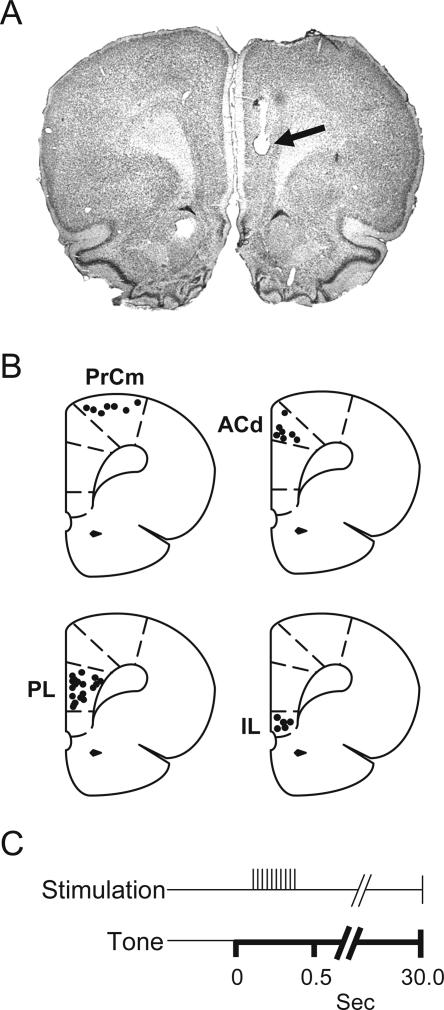
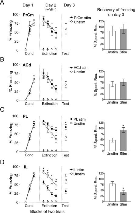
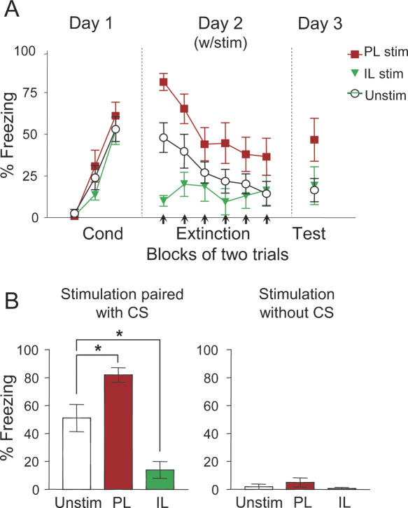
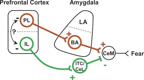
Recent studies using lesion, infusion, and unit-recording techniques suggest that the infralimbic (IL) subregion of medial prefrontal cortex (mPFC) is necessary for the inhibition of conditioned fear following extinction. Brief microstimulation of IL paired with conditioned tones, designed to mimic neuronal tone responses, reduces the expression of conditioned fear to the tone. In the present study we used microstimulation to investigate the role of additional mPFC subregions: the prelimbic (PL), dorsal anterior cingulate (ACd), and medial precentral (PrCm) cortices in the expression and extinction of conditioned fear. These are tone-responsive areas that have been implicated in both acquisition and extinction of conditioned fear. In contrast to IL, microstimulation of PL increased the expression of conditioned fear and prevented extinction. Microstimulation of ACd and PrCm had no effect. Under low-footshock conditions (to avoid ceiling levels of freezing), microstimulation of PL and IL had opposite effects, respectively increasing and decreasing freezing to the conditioned tone. We suggest that PL excites amygdala output and IL inhibits amygdala output, providing a mechanism for bidirectional modulation of fear expression.


Figures

Figure 1.
Figure 1.
Placements of stimulating electrodes in different prefrontal subregions. (A) Photomicrograph showing the tip of a stimulating electrode in PL (arrow). (B) Coronal drawings (bregma +3.20 mm) showing the location of stimulating electrodes in PrCm, ACd, PL, and IL. (C) A brief train of pulses was delivered to PrCm, ACd, PL, or IL 100–400 msec after tone onset on Day 2, in order to simulate tone responses.
Figure 2.
Figure 2.
PL microstimulation, but not PrCm or ACd, impairs extinction. (A) Microstimulation of PrCm (n = 7) or (B) ACd (n = 7) on Day 2 had no effect on acquisition of extinction or subsequent retrieval of extinction (Day 3) when compared with Unstim group (n = 7; n = 9, respectively). (C) PL microstimulation (n = 11) showed a trend of having a slower rate of extinction on Day 2 when compared with Unstim group (n = 9; P = 0.09). Furthermore, animals that received PL microstimulation on Day 2 had significantly greater recovery of freezing when compared with the Unstim group (Figs. in the right; P < 0.01). (D) IL microstimulation data from Milad et al. (2004) is shown for comparison purposes.
Figure 3.
Figure 3.
Under low footshock conditions, PL microstimulation increases fear expression, whereas IL microstimulation decreases fear expression. (A) On Day 2, PL microstimulation (n = 8) increased conditioned freezing whereas IL microstimulation (n = 6) decreased freezing, compared with the Unstim group (n = 12). On Day 3, there was no significant difference between groups (ANOVA, P = 0.08). (B, left) Bar graph showing percent freezing for the very first extinction trial on Day 2 (one-way ANOVA P < 0.001; Unstim vs. IL: P = 0.020; Unstim vs. PL: P = 0.036; IL vs. PL: P < 0.001). (Right) Microstimulation delivered in the absence of conditioned tone had no effect. CS = conditioned stimulus (tone).
Figure 4.
Figure 4.
Model of prelimbic (PL) and infralimbic (IL) interactions with the amygdala. During PL microstimulation, feedforward excitation activates (plus sign) the basal amygdala (BA) which in turn activates the medial division of the central nucleus (CeM) to produce conditioned fear responses. In contrast, IL microstimulation excites GABAergic cells in the intercalated (ITC) region as well as the lateral division of the central nucleus (CeL). Excitation of both of these nuclei inhibits (minus sign) the output of CeM and reduces fear. Future experiments will determine if local inhibitory interactions between PL and IL (arrow) are important for controlling fear expression.

Similar articles

Cited by

References

    1. Akirav I., Raizel H., Maroun M. Enhancement of conditioned fear extinction by infusion of the GABA agonist muscimol into the rat prefrontal cortex and amygdala. Eur. J. Neurosci. 2006;23:758–764. - PubMed
    1. Amat J., Baratta M.V., Paul E., Bland S.T., Watkins L.R., Maier S.F. Medial prefrontal cortex determines how stressor controllability affects behavior and dorsal raphe nucleus. Nat. Neurosci. 2005;8:365–371. - PubMed
    1. Anglada-Figueroa D., Quirk G.J. Lesions of the basal amygdala block expression of conditioned fear but not extinction. J. Neurosci. 2005;25:9680–9685. - PMC - PubMed
    1. Baeg E.H., Kim Y.B., Jang J., Kim H.T., Mook-Jung I., Jung M.W. Fast spiking and regular spiking neural correlates of fear conditioning in the medial prefrontal cortex of the rat. Cereb. Cortex. 2001;11:441–451. - PubMed
    1. Barrett D., Shumake J., Jones D., Gonzalez-Lima F. Metabolic mapping of mouse brain activity after extinction of a conditioned emotional response. J. Neurosci. 2003;23:5740–5749. - PMC - PubMed

Publication types

LinkOut - more resources