. 2006 Dec;16(12):1465-79.
doi: 10.1101/gr.5460106.

Novel patterns of genome rearrangement and their association with survival in breast cancer

Affiliations

Novel patterns of genome rearrangement and their association with survival in breast cancer

James Hicks et al. Genome Res. 2006 Dec.

Abstract

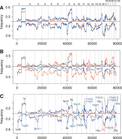
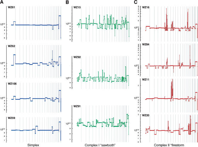
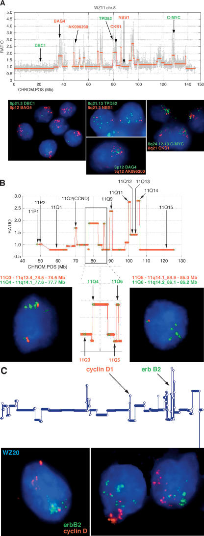
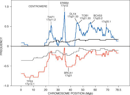
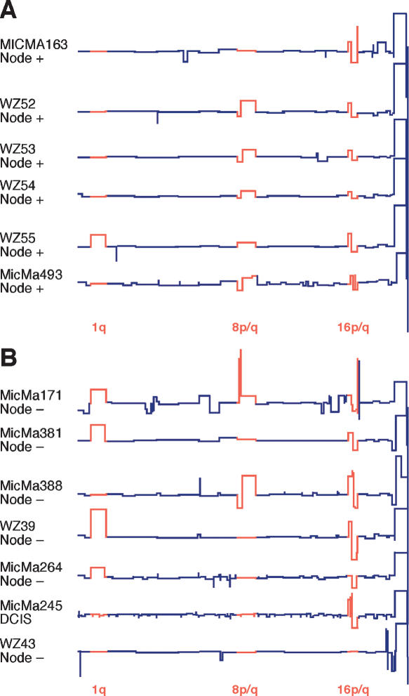
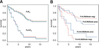
Representational Oligonucleotide Microarray Analysis (ROMA) detects genomic amplifications and deletions with boundaries defined at a resolution of approximately 50 kb. We have used this technique to examine 243 breast tumors from two separate studies for which detailed clinical data were available. The very high resolution of this technology has enabled us to identify three characteristic patterns of genomic copy number variation in diploid tumors and to measure correlations with patient survival. One of these patterns is characterized by multiple closely spaced amplicons, or "firestorms," limited to single chromosome arms. These multiple amplifications are highly correlated with aggressive disease and poor survival even when the rest of the genome is relatively quiet. Analysis of a selected subset of clinical material suggests that a simple genomic calculation, based on the number and proximity of genomic alterations, correlates with life-table estimates of the probability of overall survival in patients with primary breast cancer. Based on this sample, we generate the working hypothesis that copy number profiling might provide information useful in making clinical decisions, especially regarding the use or not of systemic therapies (hormonal therapy, chemotherapy), in the management of operable primary breast cancer with ostensibly good prognosis, for example, small, node-negative, hormone-receptor-positive diploid cases.


Figures

Figure 1.
Figure 1.
Comparative frequency plots of amplification (up) and deletion (down) in various data sets. Frequency calculated on normalized, segmented ROMA profiles using a minimum of six consecutive probes identifying a segment with a minimum mean of 0.1 above (amplification) or below (deletion) baseline. Frequencies are plotted only for chromosomes 1–22. (A) Total Swedish data set (red) versus total Norwegian data set (blue). (B) Swedish diploid subset (blue) versus total Swedish aneuploid subset (red). (C) Swedish diploid 7-yr survivors (red) versus Swedish diploid 7-yr nonsurvivors (blue).
Figure 2.
Figure 2.
Major types of tumor genomic profiles. Segmentation profiles for individual tumors representing each category: (A) simplex; (B) complex type I or sawtooth; (C) complex type II or firestorm. Scored events consist of a minimum of six consecutive probes in the same state. The y-axis displays the geometric mean value of two experiments on a log scale. Note that the scale of the amplifications in C is compressed relative to A and B owing to the high levels of amplification in firestorms. Chromosomes 1–22 plus X and Y are displayed in order from left to right according to probe position.
Figure 3.
Figure 3.
Validation of peaks and valleys in ROMA profiles by interphase FISH. (A) Expanded ROMA profile of a firestorm on chromosome 8 in the diploid tumor WZ11. The graph shows the normalized raw data (gray) and segmented profile (red) along with the genes for which the probes shown in the FISH images were constructed. Several distinct conditions are exemplified in the images. First, the ROMA profile indicates that the 8p arm is deleted distal to the 8p12 cytoband yielding a single copy of DBC1 (green), but >10 tightly clustered copies of BAG4, which is located in the frequently amplified 8p12 locus (Garcia et al. 2005). Tight clusters of multiple copies corresponding to ROMA peaks are also shown in the FISH images for CKS1A, MYC, TPD52, and the uncharacterized ORF AK096200. Note that the FISH signals corresponding to distinct loci cluster together irrespective of their distance on the same arm (CKS1A/MYC) or across the centromere (BAG4/AK096200). Finally, the spaces between ROMA peaks on 8q, exemplified by NBN (formerly known as NBS1), uniformly show two copies as indicated by the ROMA profile. (B) Expanded view of the centromere and 11q arm from diploid tumor WZ17 showing correspondence of the copy number as measured by FISH with the copy number predicted by the ROMA profile. The y-axis represents the segmented ratios of sample versus control. Chromosome position on the x-axis is in megabases according to Freeze 15 (April 2003) on the UCSC Genome Browser (Karolchik et al. 2003). FISH probes were amplified from primers identified from specific loci using PROBER software (Methods).The insert outlined in black is magnified to show specific details. Comparative data for the probes shown in black are not shown but are available on our Web site. In the boxed region, note that in the nonamplified regions the ROMA profile predicts two copies of the arm proximal to the leftmost amplification. Consistent with the profile, the FISH image shows two copies of probe 11Q3, with one of the spots located in the cluster along with the amplified copies. The amplicon to the right yields four copies by FISH (probe 11Q4). The ROMA profile for the amplicon represented by probe 11Q6 suggests that it is in a region in which the surrounding nonamplified portion of the arm is deleted. This arrangement is commonly observed in firestorms and is confirmed by the FISH image showing one pair of the loci 11q5 and 11Q6 together, representing the intact arm, and no copy of probe 11Q5 in the amplified cluster of spots for 11Q6. (C) Profile of tumor WZ19 in which two firestorms are observed on chromosomes 11q and 17q. In contrast to the overlapping clusters shown in A, amplifications on unrelated arms visualized using FISH probes for CCND1 and ERBB2 cluster independently in the nucleus.
Figure 4.
Figure 4.
Frequency plots of amplification and deletions in tumors containing clustered amplifications (firestorms) on chromosome 17. Lines represent histograms of the number of events for each probe in segmented ROMA profiles over threshold as in Figure 1 for two subsets extracted from the combined Scandinavian data set. Blue and red lines represent amplifications and deletions, respectively, in the subset of 23 tumors containing firestorms on chromosome 17, each showing clear peaks (valleys) of activity. Black and gray lines represent equivalent events in a set of 53 tumors in which firestorms are not observed on chromosome 17.
Figure 5.
Figure 5.
Comparison of Grade I and DCIS tumors by ROMA. Segmented ROMA profiles of six node-positive (Fig. 5A) and seven node-negative (Fig. 5B) Grade I or DCIS tumors, representing a total of 24 examples from the combined Swedish and Norwegian collections. Most frequent rearrangements are depicted in red.
Figure 6.
Figure 6.
Kaplan-Meier plots of the Swedish diploid subset grouped according to the Firestorm Index (F). (A) Complete Swedish diploid data set grouped according to three different discriminator settings (Fd) of F: Fd = 0.08 (red); Fd = 0.09 (blue); Fd = 0.1 (green). (B) Swedish diploid data set separated into node-negative (red) and node-positive (blue) subsets with Fd set to 0.09.

Similar articles

Cited by

References

    1. Ahr A., Karn T., Solbach C., Seiter T., Strebhardt K., Holtrich U., Kaufmann M., Karn T., Solbach C., Seiter T., Strebhardt K., Holtrich U., Kaufmann M., Solbach C., Seiter T., Strebhardt K., Holtrich U., Kaufmann M., Seiter T., Strebhardt K., Holtrich U., Kaufmann M., Strebhardt K., Holtrich U., Kaufmann M., Holtrich U., Kaufmann M., Kaufmann M. Identification of high risk breast-cancer patients by gene expression profiling. Lancet. 2002;359:131–132. - PubMed
    1. Albertson D.G. Profiling breast cancer by array CGH. Breast Cancer Res. Treat. 2003;78:289–298. - PubMed
    1. Al Kuraya K., Schraml P., Torhorst J., Tapia C., Zaharieva B., Novotny H., Spichtin H., Maurer R., Mirlacher M., Kochli O., Schraml P., Torhorst J., Tapia C., Zaharieva B., Novotny H., Spichtin H., Maurer R., Mirlacher M., Kochli O., Torhorst J., Tapia C., Zaharieva B., Novotny H., Spichtin H., Maurer R., Mirlacher M., Kochli O., Tapia C., Zaharieva B., Novotny H., Spichtin H., Maurer R., Mirlacher M., Kochli O., Zaharieva B., Novotny H., Spichtin H., Maurer R., Mirlacher M., Kochli O., Novotny H., Spichtin H., Maurer R., Mirlacher M., Kochli O., Spichtin H., Maurer R., Mirlacher M., Kochli O., Maurer R., Mirlacher M., Kochli O., Mirlacher M., Kochli O., Kochli O., et al. Prognostic relevance of gene amplifications and coamplifications in breast cancer. Cancer Res. 2004;64:8534–8540. - PubMed
    1. Balmain A., Gray J., Ponder B., Gray J., Ponder B., Ponder B. The genetics and genomics of cancer. Nat. Genet. 2003;33:238–244. - PubMed
    1. Berns E.M., de Klein A., van Putten W.L., van Staveren I.L., Bootsma A., Klijn J.G., Foekens J.A., de Klein A., van Putten W.L., van Staveren I.L., Bootsma A., Klijn J.G., Foekens J.A., van Putten W.L., van Staveren I.L., Bootsma A., Klijn J.G., Foekens J.A., van Staveren I.L., Bootsma A., Klijn J.G., Foekens J.A., Bootsma A., Klijn J.G., Foekens J.A., Klijn J.G., Foekens J.A., Foekens J.A. Association between RB-1 gene alterations and factors of favourable prognosis in human breast cancer, without effect on survival. Int. J. Cancer. 1995;64:140–145. - PubMed

Publication types