

Similarity and differences in the Lactobacillus acidophilus group identified by polyphasic analysis and comparative genomics
- PMID: 17142402
- PMCID: PMC1797336
- DOI: 10.1128/JB.01393-06
Similarity and differences in the Lactobacillus acidophilus group identified by polyphasic analysis and comparative genomics
Abstract
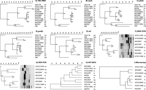
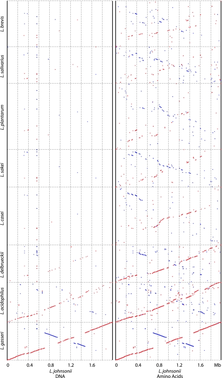
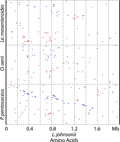
A set of lactobacilli were investigated by polyphasic analysis. Multilocus sequence analysis, DNA typing, microarray analysis, and in silico whole-genome alignments provided a remarkably consistent pattern of similarity within the Lactobacillus acidophilus complex. On microarray analysis, 17 and 5% of the genes from Lactobacillus johnsonii strain NCC533 represented variable and strain-specific genes, respectively, when tested against four independent isolates of L. johnsonii. When projected on the NCC533 genome map, about 10 large clusters of variable genes were identified, and they were enriched around the terminus of replication. A quarter of the variable genes and two-thirds of the strain-specific genes were associated with mobile DNA. Signatures for horizontal gene transfer and modular evolution were found in prophages and in DNA from the exopolysaccharide biosynthesis cluster. On microarray hybridizations, Lactobacillus gasseri strains showed a shift to significantly lower fluorescence intensities than the L. johnsonii test strains, and only genes encoding very conserved cellular functions from L. acidophilus hybridized to the L. johnsonii array. In-silico comparative genomics showed extensive protein sequence similarity and genome synteny of L. johnsonii with L. gasseri, L. acidophilus, and Lactobacillus delbrueckii; moderate synteny with Lactobacillus casei; and scattered X-type sharing of protein sequence identity with the other sequenced lactobacilli. The observation of a stepwise decrease in similarity between the members of the L. acidophilus group suggests a strong element of vertical evolution in a natural phylogenetic group. Modern whole-genome-based techniques are thus a useful adjunct to the clarification of taxonomical relationships in problematic bacterial groups.
Figures


References
-
- Altermann, E., W. M. Russell, M. A. Azcarate-Peril, R. Barrangou, B. L. Buck, O. McAuliffe, N. Souther, A. Dobson, T. Duong, M. Callanan, S. Lick, A. Hamrick, R. Cano, and T. R. Klaenhammer. 2005. Complete genome sequence of the probiotic lactic acid bacterium Lactobacillus acidophilus NCFM. Proc. Natl. Acad. Sci. USA 102:3906-3912. - PMC - PubMed
-
- Altschul, S. F., W. Gish, W. Miller, E. W. Myers, and D. J. Lipman. 1990. Basic local alignment search tool. J. Mol. Biol. 215:403-410. - PubMed
-
- Bapteste, E., Y. Boucher, J. Leigh, and W. F. Doolittle. 2004. Phylogenetic reconstruction and lateral gene transfer. Trends Microbiol. 12:406-411. - PubMed
-
- Bernardeau, M., M. Guguen, and J. P. Vernoux. 2006. Beneficial lactobacilli in food and feed: long-term use, biodiversity and proposals for specific and realistic safety assessments. FEMS Microbiol. Rev. 30:487-513. - PubMed
-
- Boekhorst, J., R. J. Siezen, M. C. Zwahlen, D. Vilanova, R. D. Pridmore, A. Mercenier, M. Kleerebezem, W. M. de Vos, H. Brussow, and F. Desiere. 2004. The complete genomes of Lactobacillus plantarum and Lactobacillus johnsonii reveal extensive differences in chromosome organization and gene content. Microbiology 150:3601-3611. - PubMed
Publication types
MeSH terms
Substances
LinkOut - more resources
Full Text Sources
Other Literature Sources
Molecular Biology Databases
