. 2007 Jan 26;282(4):2450-5.
doi: 10.1074/jbc.C600286200. Epub 2006 Dec 1.

Proteome-wide analysis in Saccharomyces cerevisiae identifies several PHD fingers as novel direct and selective binding modules of histone H3 methylated at either lysine 4 or lysine 36

Affiliations

Proteome-wide analysis in Saccharomyces cerevisiae identifies several PHD fingers as novel direct and selective binding modules of histone H3 methylated at either lysine 4 or lysine 36

Xiaobing Shi et al. J Biol Chem. .

Abstract

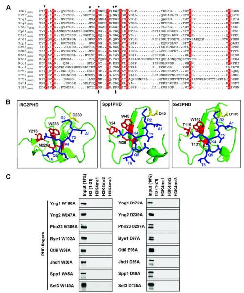
The PHD finger motif is a signature chromatin-associated motif that is found throughout eukaryotic proteomes. Here we have determined the histone methyl-lysine binding activity of the PHD fingers present within the Saccharomyces cerevisiae proteome. We provide evidence on the genomic scale that PHD fingers constitute a general class of effector modules for histone H3 trimethylated at lysine 4 (H3K4me3) and histone H3 trimethylated at lysine 36 (H3K36me3). Structural modeling of PHD fingers demonstrates a conserved mechanism for recognizing the trimethyl moiety and provides insight into the molecular basis of affinity for the different methyl-histone ligands. Together, our study suggests that a common function for PHD fingers is to transduce methyl-lysine events and sheds light on how a single histone modification can be linked to multiple biological outcomes.


Figures

FIGURE 1
FIGURE 1. Identification of histone methyl-lysine binding activity for the PHD fingers present within the S. cerevisiae proteome
A, peptide microarrays identifies the S. cerevisiae PHD fingers with H3K4me binding activity. The indicated biotinylated histone peptides were arrayed in triplicate onto streptavidin-coated slides as shown in the schematic and probed with the indicated GST-PHD finger fusion proteins. ING2(PHD) and GST are shown as positive and negative controls, respectively. B, H3K4me3 peptides bind to PHD fingers printed on protein microarray chips. The indicated GST-PHD finger fusion proteins were arrayed in duplicate onto nitrocellulose slides as shown in the schematic and probed with the indicated histone peptide. Anti-GST antibody probe is shown as a control for protein loading.
FIGURE 2
FIGURE 2. H3K4me3 or H3K36me3 recognition is a common property of S. cerevisiae PHD fingers
A, preferential binding to higher states of H3K4 methylation by S. cerevisiae PHD fingers. Western analysis of histone peptide pulldowns under stringent conditions (300 mM NaCl) with the indicated GST fusion proteins and biotinylated peptides (aa: amino acids). B, a number of S. cerevisiae PHD fingers bind to H3K36me3. The indicated proteins were tested in pulldown assays as described for A, except 150 mM NaCl binding buffer was utilized. C, disassociation constants (Kd) of the indicated PHD fingers with the indicated peptide were determined by Trp fluorescence.
FIGURE 3
FIGURE 3. Molecular features of H3K4me3 binding by S. cerevisiae PHD fingers
A, sequence alignment of PHD fingers from S. cerevisiae with those from human ING2 and BPTF. Zinc-coordinating residues are shaded in red; residues in the ING2 PHD finger essential for interaction of methyl-H3K4 (triangles) and H3R2 (circles) are indicated. Note that BPTF(PHD) utilizes an extra tyrosine to form a K4me-binding cage (18). Residues substituted for the mutagenesis studies are indicated with arrows at the bottom of the alignment. Sequence alignment was produced with the ESPript web server. B, structural models of Spp1(PHD) and Set3(PHD) complexed with H3(1–6)K4me3 peptide. Blue, histone peptide; red, residues that form methyl-K4 binding cage; yellow, residues essential for H3R2 interaction. Structure of the ING2(PHD)-H3K4me3 complex is shown for comparison. C, peptide pulldown assay with the indicated PHD finger mutants (left, W>A; right, D>A) performed as described for Fig. 2A.
FIGURE 4
FIGURE 4. Molecular features of H3K36me3-binding by S. cerevisiae PHD fingers
A, structural models of Ecm5(PHD) in complex with H3(1–6)K4me3 (right panel) and H3(33–38)K36me3 (left panel) peptides as in Fig. 3B, except that the yellow residue indicates Glu1254 (see “Results and Discussion”). B, structural model of Nto1(PHD), residues that form the aromatic cage are indicated. Residue Phe285 is shown in yellow (see “Results and Discussion”). C, peptide pulldown assay with the indicated GST-fused PHD mutants as described for Fig. 2B.

Similar articles

Cited by

References

    1. Turner BM. BioEssays. 2000;22:836–845. - PubMed
    1. Strahl BD, Allis CD. Nature. 2000;403:41–45. - PubMed
    1. Jenuwein T, Allis CD. Science. 2001;293:1074–1080. - PubMed
    1. de la Cruz X, Lois S, Sanchez-Molina S, Martinez-Balbas MA. BioEssays. 2005;27:164–175. - PubMed
    1. Daniel JA, Pray-Grant MG, Grant PA. Cell Cycle. 2005;4:919–926. - PubMed

Publication types