. 2007 Feb 16;282(7):5026-5036.
doi: 10.1074/jbc.M609360200. Epub 2006 Dec 1.

Essential role for Co-chaperone Fkbp52 but not Fkbp51 in androgen receptor-mediated signaling and physiology

Affiliations

Essential role for Co-chaperone Fkbp52 but not Fkbp51 in androgen receptor-mediated signaling and physiology

Weidong Yong et al. J Biol Chem. .

Abstract

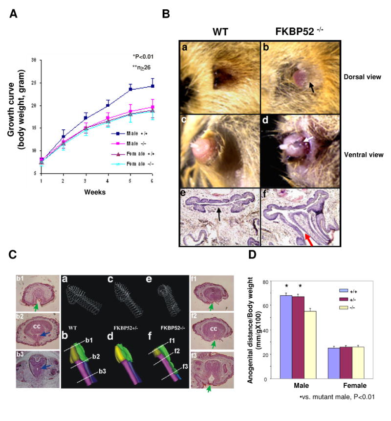
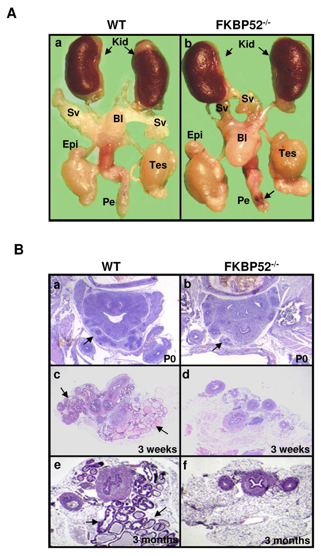
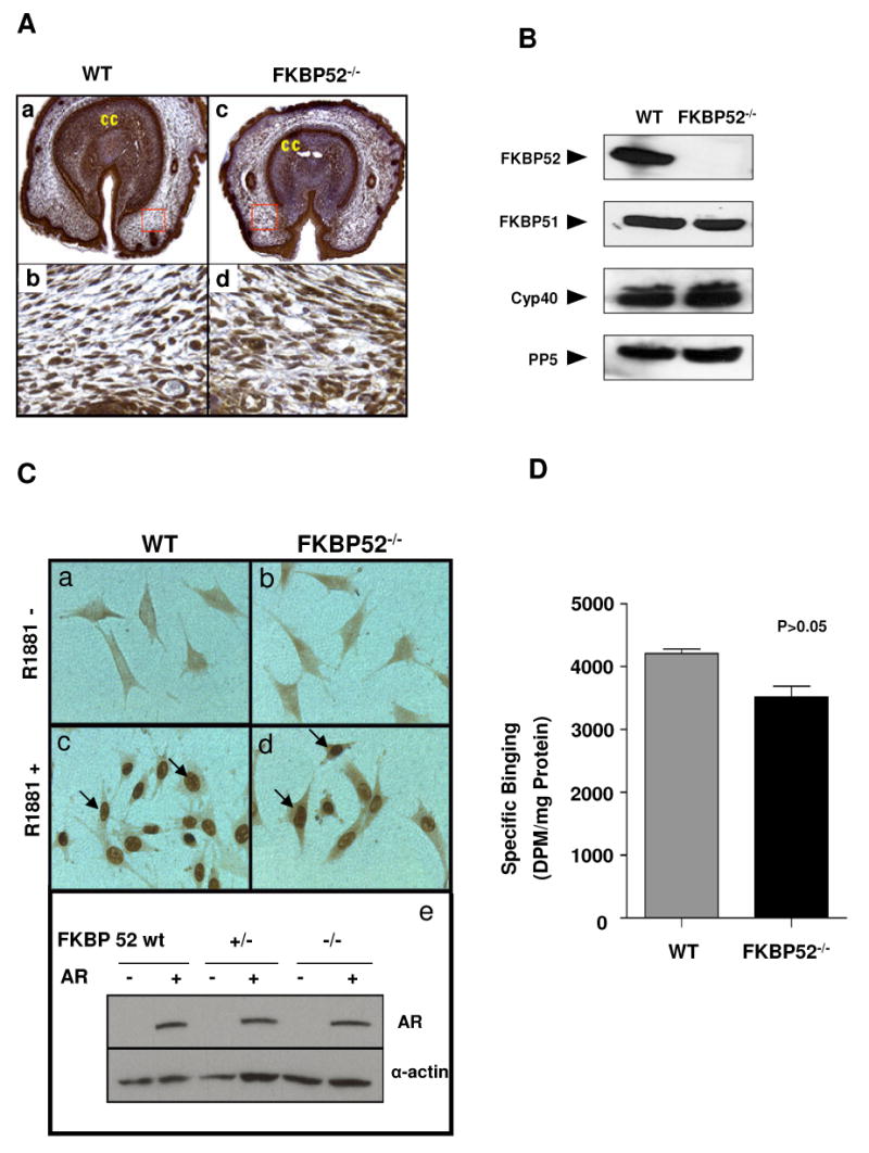
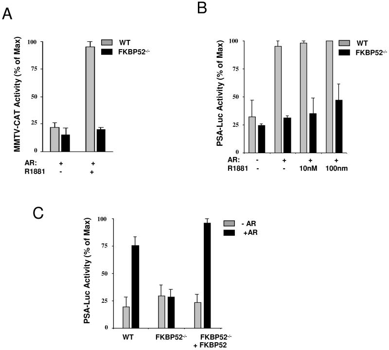
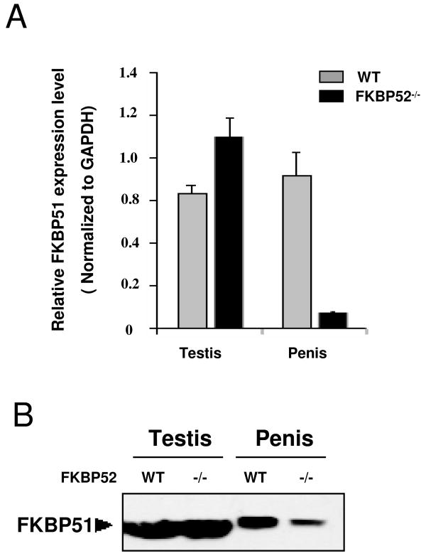
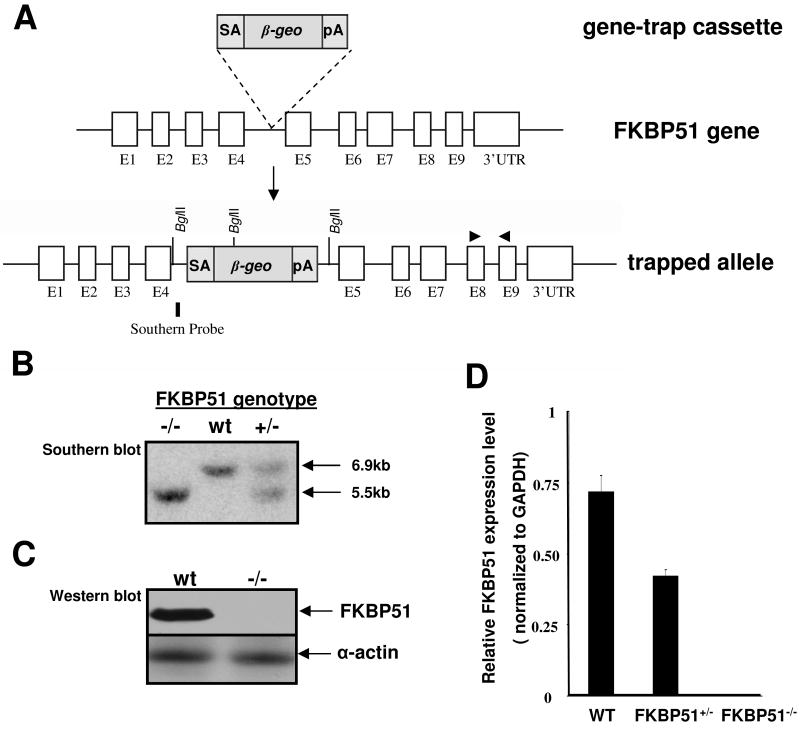
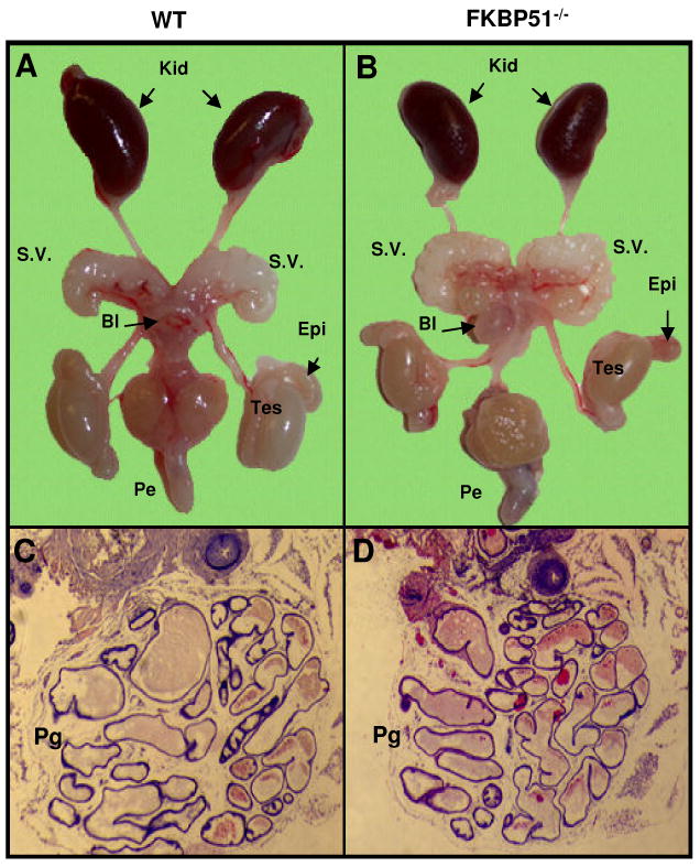
Fkbp52 and Fkbp51 are tetratricopeptide repeat proteins found in steroid receptor complexes, and Fkbp51 is an androgen receptor (AR) target gene. Although in vitro studies suggest that Fkbp52 and Fkbp51 regulate hormone binding and/or subcellular trafficking of receptors, the roles of Fkbp52 and Fkbp51 in vivo have not been extensively investigated. Here, we evaluate their physiological roles in Fkbp52-deficient and Fkbp51-deficient mice. Fkbp52-deficient males developed defects in select reproductive organs (e.g. penile hypospadias and prostate dysgenesis but normal testis), pointing to a role for Fkbp52 in AR-mediated signaling and function. Surprisingly, ablation of Fkbp52 did not affect AR hormone binding or nuclear translocation in vivo and in vitro. Molecular studies in mouse embryonic fibroblast cells uncovered that Fkbp52 is critical to AR transcriptional activity. Interestingly, Fkbp51 expression was down-regulated in Fkbp52-deficient males but only in affected tissues, providing further evidence of tissue-specific loss of AR activity and suggesting that Fkbp51 is an AR target gene essential to penile and prostate development. However, Fkbp51-deficient mice were normal, showing no defects in AR-mediated reproductive function. Our work demonstrates that Fkbp52 but not Fkbp51 is essential to AR-mediated signaling and provides evidence for an unprecedented Fkbp52 function, direct control of steroid receptor transcriptional activity.


Figures

Figure 1
Figure 1
Morphological and histological analysis of FKBP52-deficient males. (A) Growth curves of WT and FKBP52-deficient mice; (B) Comparison of adult wild-type and FKBP52-deficient male external genitalia at dorsal (a and b) and ventral surfaces (c and d). Arrows (in b and d) indicate the under-developed foreskin and ectopic urethral opening at the ventral aspect compared to normal morphology in controls (a and c). Histological sections of wild-type (e) and FKBP52-deficient (f) male genitalia. A red arrow indicates ectopic urethral opening at the ventral aspect compared to normal morphology in controls (black arrow in e). (C) Three-dimensional reconstruction of the mouse E18.5 penises. Wire frame images of outer penile skin and urethra of the three FKBP52 genotypes are shown in a, c and e. In b, d and f, the skins had been artificially removed. The glans penis is colored in gold, while the urethral opening is green, corpus cavernosum (cc) is purple, and the closed urethra is blue. Note that the urethral opening persisted throughout the FKBP52-deficient penile shaft, while normal controls only have a temporary urethral opening at distal end. Side panels show representative sections of wild-type and mutant penises. Green arrows indicate open urethra, blue arrows indicate closed urethra. (D) Comparison of anogenital distances in wild-type, heterozygous and FKBP52-deficient mice. The anogenital distance, normalized by the animal body weight, in FKBP52-deficient males was significantly shorter compared to littermate wild-type and heterozygous males.
Figure 2
Figure 2
(A) Morphological comparison of male internal reproductive organs in wild-type (a) and FKBP52-deficient adult mice (b). FKBP52-deficient males have overall normal testis (Tes) formation and normal epididymis (Epi), but significantly smaller seminal vesicles (SV). Kid: kidney; Bl: bladder; Pe: penis; Black arrow indicates urethral opening. (B) Histological analysis of prostate gland development in FKBP52-deficient and age-matched littermate control mice at birth (P0) to 3 month old. Prostate glands are initially formed during embryonic developmental in both wild-type (a) FKBP52-deficient mutant (b), but lack further growth after the birth (d) and eventually become dysgenic in FKBP52-deficient adult males (f) compared to littermate wild-type mice (c and e). Prostate glands are indicated by black arrows.
Figure 3
Figure 3
Analysis of FKBP52 deficiency on AR nuclear translocation and hormone binding. (A) Immunochemical staining shows that AR is highly expressed in all cell types of wild-type (a) and FKBP52-deficient genital tubercles (c), and that AR nuclear localization is not altered in FKBP52-deficient mutants (d) compared to wild-type controls (b). (B) Ablation of FKBP52 has no compensatory effect on expression of FKBP51, Cyp40 and PP5 in FKBP52-deficient MEF cells. (C) Using AR-stably transfected MEF cells to determine the AR nuclear translocation activity in FKBP52-deficient cells. Without hormone (R1881) treatment (a and b), ARs are mainly localized in cytoplasm. Upon R1881 treatment, ARs in both wild-type (c) and FKBP52-deficient (d) MEF cells translocate to nuclei in a similar fashion. The overall AR expression levels in AR-transfected cells are comparable among these cell lines, as evaluated by Western blot analysis (e). Genotypes of the cells are as indicated. (D) Measurement of AR hormone-binding capacity in cytosols from AR transfected wild-type and FKBP52-deficient MEF cells using [3H]mibolerone. The results shown are the means +/- SEM of two independent experiments, each performed in triplicate. No significant effect of FKBP52 ablation is seen on AR hormone-binding function.
Figure 4
Figure 4
Analysis of AR transcriptional activity in FKBP52-deficient MEF cells. (A) Transcriptional enhancement activity by hormone at MMTV-CAT is inhibited in FKBP52-deficient cells. (B) Transcriptional enhancement activity at the PSA-luciferase reporter is also inhibited. (C) Analysis of AR transcription activity in FKBP52-deficient MEF cells with re-introduction of human FKBP52. Values represent the means +/− SEM of four independent experiments.
Figure 5
Figure 5
Analyses of FKBP51 expression in the testis and penis. (A) Quantitative RT-PCR analysis of FKBP51 in testis and penis of wild-type and FKBP52-deficient males (values represent the means +/− SEM of four independent experiments). In FKBP52-deficient penile tissues, FKBP51 is dramatically down-regulated. (B) The tissue-selectivity of FKBP51 expression was confirmed by Western-blotting.
Figure 6
Figure 6
Generation of FKBP51-deficient mice. (A) Genomic structure of the mouse FKBP51 gene, gene trap vector, and FKBP51 mutant allele. (B) Southern blot, (C) Western blot, and (D) qRT-PCR analyses confirm the FKBP51 mutant allele to be null. For Southern blot, the genomic DNA was digested by Bgl II (New England Biolabs). The probe indicated in (A) reveals a 6.9kb fragment from wild type allele and a 5.5kb fragment from FKBP51 mutant allele. Primers for qRT-PCR analysis are indicated by a pair of triangles in (A).
Figure 7
Figure 7
Morphological comparison of male internal reproductive organs in wild-type (A) and FKBP51-deficient adult mice (B). FKBP51-deficient males have overall normal testis (Tes) formation, normal epididymis (Epi) normal seminal vesicles (SV). Kid: kidney; Bl: bladder; Pe: penis; (C and D) Histological analysis of prostate glands in adult FKBP51-deficient and age-matched littermate control mice. FKBP51 mutant adult males have normal prostate glands (Pg).

References

    1. Tsai MJ, O'Malley BW. Annual review of biochemistry. 1994;63:451–486. - PubMed
    1. Beato M, Klug J. Hum Reprod Update. 2000;6(3):225–236. - PubMed
    1. Danielian PS, White R, Lees JA, Parker MG. The EMBO journal. 1992;11(3):1025–1033. - PMC - PubMed
    1. Mangelsdorf DJ, Thummel C, Beato M, Herrlich P, Schutz G, Umesono K, Blumberg B, Kastner P, Mark M, Chambon P, Evans RM. Cell. 1995;83(6):835–839. - PMC - PubMed
    1. He B, Gampe RT, Jr, Kole AJ, Hnat AT, Stanley TB, An G, Stewart EL, Kalman RI, Minges JT, Wilson EM. Mol Cell. 2004;16(3):425–438. - PubMed

Publication types

MeSH terms