

The quaternary structure of the amidase from Geobacillus pallidus RAPc8 is revealed by its crystal packing
- PMID: 17142891
- PMCID: PMC2225364
- DOI: 10.1107/S1744309106043855
The quaternary structure of the amidase from Geobacillus pallidus RAPc8 is revealed by its crystal packing
Abstract
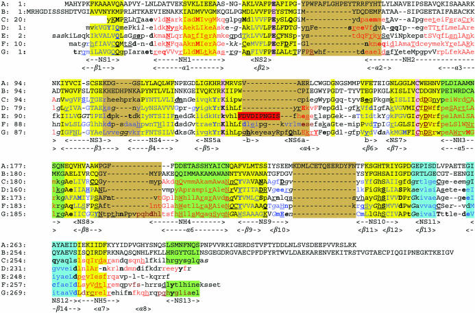
The amidase from Geobacillus pallidus RAPc8, a moderate thermophile, is a member of the nitrilase enzyme superfamily. It converts amides to the corresponding acids and ammonia and has application as an industrial catalyst. RAPc8 amidase has been cloned and functionally expressed in Escherichia coli and has been purified by heat treatment and a number of chromatographic steps. The enzyme was crystallized using the hanging-drop vapour-diffusion method. Crystals produced in the presence of 1.2 M sodium citrate, 400 mM NaCl, 100 mM sodium acetate pH 5.6 were selected for X-ray diffraction studies. A data set having acceptable statistics to 1.96 A resolution was collected under cryoconditions using an in-house X-ray source. The space group was determined to be primitive cubic P4(2)32, with unit-cell parameter a = 130.49 (+/-0.05) A. The structure was solved by molecular replacement using the backbone of the hypothetical protein PH0642 from Pyrococcus horikoshii (PDB code 1j31) with all non-identical side chains substituted with alanine as a probe. There is one subunit per asymmetric unit. The subunits are packed as trimers of dimers with D3 point-group symmetry around the threefold axis in such a way that the dimer interface seen in the homologues is preserved.
Figures


Similar articles
-
Crystallization, diffraction data collection and preliminary crystallographic analysis of hexagonal crystals of Pseudomonas aeruginosa amidase.Acta Crystallogr Sect F Struct Biol Cryst Commun. 2007 Mar 1;63(Pt 3):214-6. doi: 10.1107/S1744309107005830. Epub 2007 Feb 23. Acta Crystallogr Sect F Struct Biol Cryst Commun. 2007. PMID: 17329817 Free PMC article.
-
Structure of an aliphatic amidase from Geobacillus pallidus RAPc8.Acta Crystallogr D Biol Crystallogr. 2007 Oct;63(Pt 10):1048-58. doi: 10.1107/S090744490703836X. Epub 2007 Sep 19. Acta Crystallogr D Biol Crystallogr. 2007. PMID: 17881822
-
A novel thermostable nitrilase superfamily amidase from Geobacillus pallidus showing acyl transfer activity.Appl Microbiol Biotechnol. 2007 Jun;75(4):801-11. doi: 10.1007/s00253-007-0883-2. Epub 2007 Mar 9. Appl Microbiol Biotechnol. 2007. PMID: 17347819
-
Structural and biochemical characterization of a nitrilase from the thermophilic bacterium, Geobacillus pallidus RAPc8.Appl Microbiol Biotechnol. 2010 Sep;88(1):143-53. doi: 10.1007/s00253-010-2734-9. Epub 2010 Jul 4. Appl Microbiol Biotechnol. 2010. PMID: 20607233
-
Purification, crystallization and preliminary X-ray analysis of a thermostable glycoside hydrolase family 43 beta-xylosidase from Geobacillus thermoleovorans IT-08.Acta Crystallogr Sect F Struct Biol Cryst Commun. 2007 Nov 1;63(Pt 11):932-5. doi: 10.1107/S1744309107046015. Epub 2007 Oct 24. Acta Crystallogr Sect F Struct Biol Cryst Commun. 2007. PMID: 18007043 Free PMC article.
Cited by
-
Structural Investigations of N-carbamoylputrescine Amidohydrolase from Medicago truncatula: Insights into the Ultimate Step of Putrescine Biosynthesis in Plants.Front Plant Sci. 2016 Mar 30;7:350. doi: 10.3389/fpls.2016.00350. eCollection 2016. Front Plant Sci. 2016. PMID: 27066023 Free PMC article.
-
Crystallization, diffraction data collection and preliminary crystallographic analysis of hexagonal crystals of Pseudomonas aeruginosa amidase.Acta Crystallogr Sect F Struct Biol Cryst Commun. 2007 Mar 1;63(Pt 3):214-6. doi: 10.1107/S1744309107005830. Epub 2007 Feb 23. Acta Crystallogr Sect F Struct Biol Cryst Commun. 2007. PMID: 17329817 Free PMC article.
-
Development and application of a transcriptional sensor for detection of heterologous acrylic acid production in E. coli.Microb Cell Fact. 2019 Aug 19;18(1):139. doi: 10.1186/s12934-019-1185-y. Microb Cell Fact. 2019. PMID: 31426802 Free PMC article.
-
The mechanism of the amidases: mutating the glutamate adjacent to the catalytic triad inactivates the enzyme due to substrate mispositioning.J Biol Chem. 2013 Oct 4;288(40):28514-23. doi: 10.1074/jbc.M113.503284. Epub 2013 Aug 14. J Biol Chem. 2013. PMID: 23946488 Free PMC article.
-
Unique aliphatic amidase from a psychrotrophic and haloalkaliphilic nesterenkonia isolate.Appl Environ Microbiol. 2011 Jun;77(11):3696-702. doi: 10.1128/AEM.02726-10. Epub 2011 Apr 15. Appl Environ Microbiol. 2011. PMID: 21498772 Free PMC article.
References
-
- Ambler, R. P., Auffret, A. D. & Clarke, P. H. (1987). FEBS Lett.215, 285–290. - PubMed
-
- Banerjee, A., Sharma, R. & Banerjee, U. C. (2002). Appl. Microbiol. Biotechnol.60, 33–44. - PubMed
-
- Brenner, C. (2002). Curr. Opin. Struct. Biol.12, 775–782. - PubMed
-
- Brown, P. R., Smyth, M. J., Clarke, P. H. & Rosemeyer, M. A. (1973). Eur. J. Biochem.34, 177–187. - PubMed
-
- Cameron, R. A., Sayed, M. & Cowan, D. A. (2005). Biochim. Biophys. Acta, 1725, 35–46. - PubMed
Publication types
MeSH terms
Substances
LinkOut - more resources
Full Text Sources
