

doi: 10.1107/S1744309106044228.
Epub 2006 Nov 4.
Structure of HLA-A*1101 in complex with a hepatitis B peptide homologue
Affiliations
- PMID: 17142892
- PMCID: PMC2225367
- DOI: 10.1107/S1744309106044228
Item in Clipboard
Structure of HLA-A*1101 in complex with a hepatitis B peptide homologue
Acta Crystallogr Sect F Struct Biol Cryst Commun.
.
Abstract
A high-resolution structure of the human MHC-I molecule HLA-A*1101 is presented in which it forms a complex with a sequence homologue of a peptide that occurs naturally in hepatitis B virus DNA polymerase. The sequence of the bound peptide is AIMPARFYPK, while that of the corresponding natural peptide is LIMPARFYPK. The peptide does not make efficient use of the middle E pocket for binding, which leads to a rather superficial and exposed binding mode for the central peptide residues. Despite this, the peptide binds with high affinity (IC50 of 31 nM).
Figures
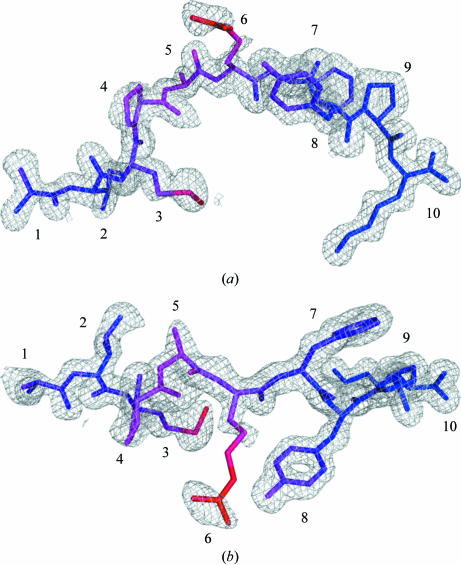
Electron density of the AIMPARFYPK peptide. The electron density is contoured at 1σ and clearly shows density for all of the peptide including the additional N-terminal alanine (left). The peptide is coloured according to B values from blue to red (13–48 Å2). (a) Side view, (b) top view. All figures were prepared with PyMOL (DeLano, 2002 ▶).
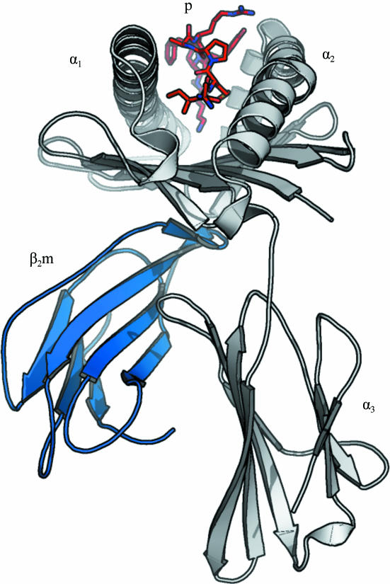
Schematic overview of the structure of HLA-A*1101 in complex with the AIMPARFYPK peptide. The AIMPARFYPK peptide (red; labelled ‘p’) is viewed from the N-terminal end. The α-chain is shown in grey with individual subdomains indicated; β2-microglobulin is in blue. Notice the relatively exposed position of the peptide in the binding groove.
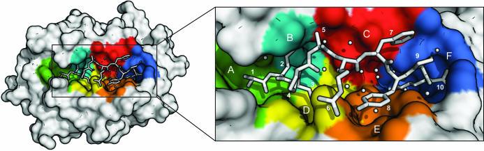
A T-cell view of the bound AIMPARFYPK peptide of the HLA-A*1101 complex. Binding pockets are indicated with colours: A, green; B, cyan; C, red; D, yellow; E, orange; F, blue. The peptide and selected water molecules suggested to be important for peptide binding are shown in white (see also Table 4 ▶). Notice the large number of water molecules beneath the peptide and that Pro4p covers Met3p.
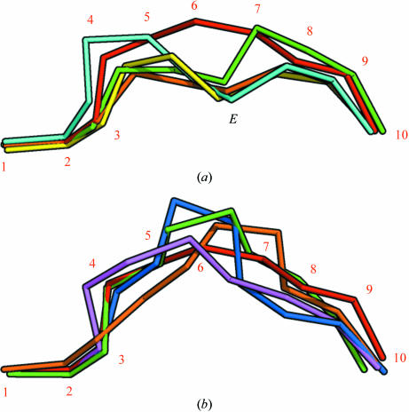
The peptide positions in the binding groove after superimposition of MHC complex structures. The Cα atoms of the peptides have been connected and α-chain residues have been removed for clarity. (a) The AIMPARFYPK peptide (red, numbered) follows a relatively superficial path, whereas other decamer peptides bound by A3 supertype molecules [green, HLA-A*6801 complex (PDB code 1tmc ; Collins et al., 1995 ▶); cyan, HLA-A*1101 complex (PDB code 1qvo ; Li & Bouvier, 2004 ▶)] achieve a deeper position by placing residue 6 in the E pocket (indicated with an E). Complexes with nonamer peptides [yellow, HLA-A*1101 complex (PDB code 1q94 ; Li & Bouvier, 2004 ▶); green, HLA-A*1101 complex (PDB code 1x7q ; Blicher et al., 2005 ▶)] utilize residue 5 for binding in the E pocket in a similar fashion. (b) Comparison of the AIMPARFYPK peptide (red) with two decamer peptides [purple, HLA-A*0201 complex (PDB code 1i4f ; Hillig et al., 2001 ▶); orange, HLA-B*2705 (PDB code 2bss ; Stewart-Jones, Di Gleria et al., 2005 ▶)] and two 11-residue peptides [blue, HLA-B*5703 (PDB code 2bvo ; Stewart-Jones, Gillespie et al., 2005 ▶); green, HLA-B*3501 (PDB code 1zsd ; Miles et al., 2005 ▶)] bound by other HLA alleles. Although the AIMPARFYPK peptide bulges out of the groove, it is not unusual for a decamer peptide. The orientation is the same as in (a).
Similar articles
-
Effect of anchor residue modifications on the stability of HLA-A11/peptide complexes.Biochem Biophys Res Commun. 1995 Jan 5;206(1):8-14. doi: 10.1006/bbrc.1995.1002. Biochem Biophys Res Commun. 1995. PMID: 7818553
-
Atomic structure of a human MHC molecule presenting an influenza virus peptide.Nature. 1992 Nov 26;360(6402):367-9. doi: 10.1038/360367a0. Nature. 1992. PMID: 1448154
-
Classical and nonclassical class I major histocompatibility complex molecules exhibit subtle conformational differences that affect binding to CD8alphaalpha.J Biol Chem. 2000 May 19;275(20):15232-8. doi: 10.1074/jbc.275.20.15232. J Biol Chem. 2000. PMID: 10809759
-
Outsize peptides bulge out of the groove.Immunol Today. 1993 Feb;14(2):51-2. doi: 10.1016/0167-5699(93)90057-r. Immunol Today. 1993. PMID: 8447932 Review.
-
Gorillas with spondyloarthropathies express an MHC class I molecule with only limited sequence similarity to HLA-B27 that binds peptides with arginine at P2.J Immunol. 2001 Mar 1;166(5):3334-44. doi: 10.4049/jimmunol.166.5.3334. J Immunol. 2001. PMID: 11207289 Review.
Cited by
-
Structural and functional distinctiveness of HLA-A2 allelic variants.Immunol Res. 2012 Sep;53(1-3):182-90. doi: 10.1007/s12026-012-8295-5. Immunol Res. 2012. PMID: 22434516 Review.
-
Humidity control as a strategy for lattice optimization applied to crystals of HLA-A*1101 complexed with variant peptides from dengue virus.Acta Crystallogr Sect F Struct Biol Cryst Commun. 2007 May 1;63(Pt 5):386-92. doi: 10.1107/S1744309107013693. Epub 2007 Apr 6. Acta Crystallogr Sect F Struct Biol Cryst Commun. 2007. PMID: 17565177 Free PMC article.
-
Structure of a SARS coronavirus-derived peptide bound to the human major histocompatibility complex class I molecule HLA-B*1501.Acta Crystallogr Sect F Struct Biol Cryst Commun. 2008 Jun 1;64(Pt 6):459-62. doi: 10.1107/S1744309108012396. Epub 2008 May 17. Acta Crystallogr Sect F Struct Biol Cryst Commun. 2008. PMID: 18540051 Free PMC article.
References
-
- Achour, A., Begue, B., Gomard, E., Paul, P., Sayagh, B., Van Pel, A. & Levy, J. P. (1986). Eur. J. Immunol.16, 597–604. - PubMed
-
- Bodmer, J., Cambon-Thomsen, A., Hors, J., Piazza, A. & Sanchez-Mazas, A. (1999). In Proceedings of the Twelfth International Histocompatibility Workshop and Conference, edited by D. Charron. Paris: EDK.
-
- Burrows, S. R., Rossjohn, J. & McCluskey, J. (2006). Trends Immunol.27, 11–16. - PubMed
-
- Buus, S., Stryhn, A., Winther, K., Kirkby, N. & Pedersen, L. O. (1995). Biochim. Biophys. Acta, 1243, 453–460. - PubMed
Publication types
MeSH terms
Substances
Associated data
- Actions
- Actions
Grants and funding
LinkOut - more resources
Full Text Sources
Other Literature Sources
Molecular Biology Databases
Research Materials
