. 2006 Dec;116(12):3277-91.
doi: 10.1172/JCI29314.

Thrombospondins deployed by thrombopoietic cells determine angiogenic switch and extent of revascularization

Affiliations

Thrombospondins deployed by thrombopoietic cells determine angiogenic switch and extent of revascularization

Hans-Georg Kopp et al. J Clin Invest. 2006 Dec.

Abstract

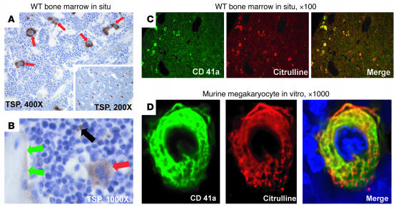
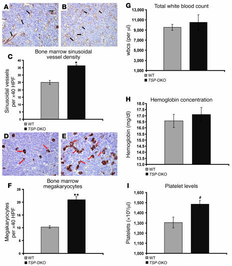
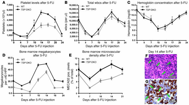
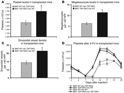
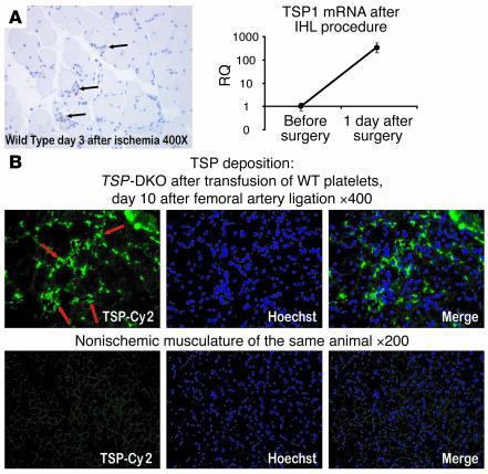
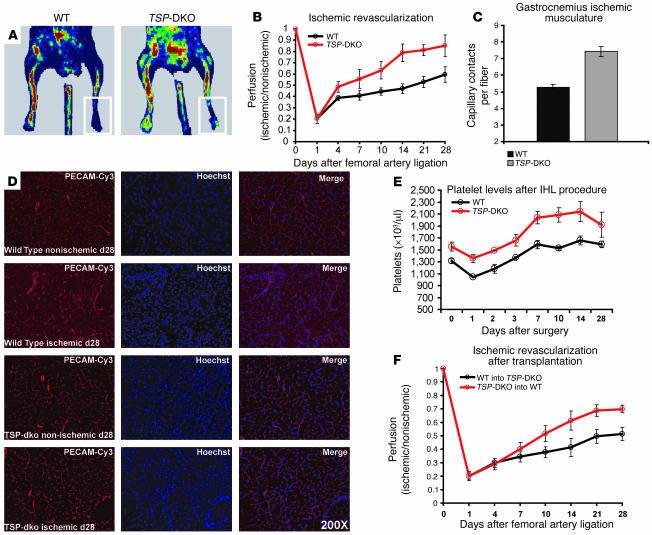
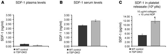
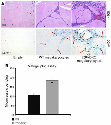
Thrombopoietic cells may differentially promote or inhibit tissue vascularization by releasing both pro- and antiangiogenic factors. However, the molecular determinants controlling the angiogenic phenotype of thrombopoietic cells remain unknown. Here, we show that expression and release of thrombospondins (TSPs) by megakaryocytes and platelets function as a major antiangiogenic switch. TSPs inhibited thrombopoiesis, diminished bone marrow microvascular reconstruction following myelosuppression, and limited the extent of revascularization in a model of hind limb ischemia. We demonstrate that thrombopoietic recovery following myelosuppression was significantly enhanced in mice deficient in both TSP1 and TSP2 (TSP-DKO mice) in comparison with WT mice. Megakaryocyte and platelet levels in TSP-DKO mice were rapidly restored, thereby accelerating revascularization of myelosuppressed bone marrow and ischemic hind limbs. In addition, thrombopoietic cells derived from TSP-DKO mice were more effective in supporting neoangiogenesis in Matrigel plugs. The proangiogenic activity of TSP-DKO thrombopoietic cells was mediated through activation of MMP-9 and enhanced release of stromal cell-derived factor 1. Thus, TSP-deficient thrombopoietic cells function as proangiogenic agents, accelerating hemangiogenesis within the marrow and revascularization of ischemic hind limbs. As such, interference with the release of cellular stores of TSPs may be clinically effective in augmenting neoangiogenesis.


Figures

Figure 1
Figure 1. In the bone marrow, TSP1 is expressed in megakaryocytes and platelets and on endosteal surfaces.
An antibody raised against citrullinated proteins results in robust and specific megakaryocyte staining. (A) Megakaryocytes (red arrows) are highly immunoreactive for TSPs. Representative images of WT bone marrow at steady state are shown. Sections were stained for TSPs. Original magnification, ×400. Inset provides a lower magnification overview of WT bone marrow; original magnification, ×200. Sections shown in A and B were counterstained with hematoxylin. (B) In addition, TSP immunoreactivity was demonstrated on endosteal osseous surfaces (green arrows) and platelets (black arrow). This would be in line with previous reports in which TSP1 has been shown to be a cytoadhesive molecule for hematopoietic stem cells. On the other hand, hematopoietic cells other than megakaryocytes and platelets did not stain with the same antibody. Frozen bone marrow section at steady state is shown. Original magnification, ×1,000. (C) WT murine steady state bone marrow was stained with an antibody against CD41 and the anti-citrulline antibody. While anti-CD41 binding was visualized with a Cy2-labeled secondary antibody (green), the anti-citrulline antibody was detected with a Cy3-labeled secondary antibody (red). It is obvious that both antibodies bind to megakaryocytes. However, the anti-citrulline antibody results in more robust and stronger staining results. Original magnification, ×100. (D) Murine megakaryocytes differentiated in culture costained with anti-CD41a and anti-citrulline as above. The merged view shows that both antibodies bind to megakaryocytes. Cytospin from in vitro–differentiated lineage negative murine bone marrow cells is shown. Original magnification, ×1,000.
Figure 2
Figure 2. TSP-DKO mice display increased bone marrow microvascular and megakaryocyte density as well as higher peripheral platelet levels compared with WT mice.
(A and B) Marrow sinusoidal microvasculature was quantified in WT (A) and TSP-DKO (B) mice after immunostaining against panendothelial cell antigen clone MECA32. Note that absolute number of cross-sectioned sinusoids (black arrows) is higher in TSP-DKO animals. Representative marrow sections at steady state are shown at original magnification, ×400. DAB was counterstained with hematoxylin. (C) TSP-DKO marrow has a higher sinusoidal microvascular density than WT marrow: 36 ± 0.7 versus 25 ± 1.4 sinusoidal vessels per field. Scored at original magnification, ×400. HPF, high-power field. *P < 0.005. (D) WT marrow, stained for TSPs. Note that only megakaryocytes and platelets are stained. Red arrows indicate differentiated, multinucleated megakaryocytes. Original magnification, ×400. DAB was counterstained with hematoxylin. (E) Megakaryocytes in TSP-DKO marrow are abundant at steady state and can be stained with an antibody against citrullinated proteins. This antibody stained megakaryocytes at the same level of differentiation as the TSP antibody (red arrows). Original magnification, ×400. DAB was counterstained with hematoxylin. (F) TSP-DKO megakaryocyte density is almost twice as high as that in WT marrow: 21 ± 1.2 versus 0 ± 0.5 megakaryocytes per field. Scored at original magnification, ×400. **P < 6 × 10⁻⁶. (G) Leukocyte counts at steady state (n = 6). Difference was not significant. (H) Analysis of hemoglobin concentration (n = 6) showed similar results. (I) TSP-DKO mice displayed significantly elevated platelet counts compared with WT controls: 1,495,000 ± 37,000/μl versus 1,305,000 ± 53,000/μl. #P < 0.05. n = 6.
Figure 3
Figure 3. TSP deficiency enhances megakaryocyte repopulation of the bone marrow and platelet production after myelosuppression.
(A) After 250 mg/kg of 5-FU treatment, WT mice experienced pancytopenia followed by rebound thrombocytosis. Platelets reverted to normal around day 24. TSP-DKO mice displayed a more rapid regeneration, with 8.9 × 10⁵ platelets/μl compared with 4.77 × 10⁵ platelets/μl in WT animals on day 7. Furthermore, rebound thrombocytosis was exaggerated, with platelets reaching 5.5 × 10⁶/μl compared with 2.9 × 10⁶/μl in WT controls on day 14 (n = 6, P = 0.006). *P < 0.05. (B) Leukocytes and (C) hemoglobin concentrations did not show significant differences between TSP-DKO and WT mice (n = 6). (D) TSP-DKO mice had higher numbers of megakaryocytes at all time points following 5-FU injection (P < 0.05). While megakaryocytes reached their lowest levels on day 7 in WT mice, megakaryocyte concentration returned to normal levels in TSP-DKO mice on day 7 (P < 0.0003). (E) After 5-FU injection, endothelial cell mass plummeted to about 2% of hematopoietic marrow surface area and reverted to normal around day 14. In TSP-DKO mice, the overall course of these changes was similar to that in WT mice. However, MECA32-positive surface area never decreased to less than 4.9% (day 7 after 5-FU injection; P < 0.005). (F and G) The same femoral marrow as represented in E stained with H&E (F) and anti-citrulline antibody (G). DAB was counterstained with hematoxylin. TSP-DKO marrow showed extreme megakaryocytosis at day 10 after 5-FU injection. Green arrows indicate megakaryocytes. Original magnification, ×400.
Figure 4
Figure 4. TSP-deficient thrombocytotic phenotype is transplantable.
(AC) Lethally irradiated (9.5 Gy) WT control mice were transplanted with TSP-DKO bone marrow and TSP-DKO mice were transplanted with WT bone marrow. Ninety days after transplantation, platelet counts were 1.7 ± 0.1 × 10⁶/μl in WT recipients transplanted with TSP-DKO bone marrow and 1.4 ± 0.6 106/μl in TSP-DKO recipients transplanted with WT bone marrow. P < 0.04. Platelet (A) and megakaryocyte levels (B) and vascular density (C) were determined in the transplanted mice. (D) When 5-FU was given (250 mg/kg i.v.) to the same mice, WT mice with TSP-deficient hematopoiesis displayed a faster platelet recovery than TSP-deficient mice that had been transplanted with WT bone marrow. On day 10, platelet levels were 0.18 ± 0.05 × 10⁶/μl in TSP-DKO mice with WT hematopoiesis compared with 1.6 ± 0.27 × 10⁶/μl in the WT recipients of TSP-deficient bone marrow (P < 0.03). Therefore, TSP from transplanted megakaryocytes, rather than TSP presented or secreted by nontransplanted bone marrow stromal components, may regulate megakaryopoiesis and thrombopoiesis following myelosuppression. *P < 0.05.
Figure 5
Figure 5. The vascular disrupting agent CA4P targets bone marrow microvascular sinusoids as well as megakaryopoiesis.
(A) CA4P delays platelet recovery and blocks rebound thrombocytosis. TSP-DKO mice were treated with 250 mg/kg 5-FU i.v. and either with an additional low dose of CA4P (25 mg/kg) every other day or with PBS. CA4P has not been shown to be myelosuppressive at this dose in long-term studies (data not shown). While 5-FU alone resulted in a typical rebound thrombocytosis, CA4P-treated mice reverted back to normal platelet levels later, and the rebound effect was completely abrogated. n = 5 in each group. P < 0.02 on day 13. (B) CA4P delays white blood cell recovery. Total peripheral blood white cell counts displayed a lower nadir and a delayed recovery albeit with a marked rebound effect that is rather atypical for white cells. (C) CA4P increases severity of anemia but does not delay red blood cell recovery. Hemoglobin levels were decreased to lower absolute levels when CA4P was added to the myelosuppressive regimen, but the overall time course of rbc regeneration was comparable to that in the group that received 5-FU only. (D) Histological analysis of the marrow on day 10 revealed that the repopulation of the marrow with hematopoietic cells was not significantly inhibited. However, the vascular microarchitecture was severely disturbed, resulting in areas of hemorrhage (black arrows). H&E staining was used. Original magnification, ×400. (E) Staining with the panendothelial cell marker antibody MECA32 showed disrupted, leaky microvasculature in the CA4P-treated mice (red arrows). DAB was counterstained with hematoxylin. Original magnification, ×400. (F) The same femurs as in D were stained with anti-citrulline antibody to identify megakaryocytes. Interestingly, it became evident that the vascular disruption seen with CA4P treatment resulted in a selective defect in megakaryocyte repopulation of the marrow, with a reversion of the TSP-DKO phenotype toward normal. Anti-citrulline staining with DAB was counterstained with hematoxylin. Original magnification, ×400.
Figure 6
Figure 6. TSPs are deposited perivascularly in ischemic musculature.
(A) Left: WT mice underwent ligation of the left femoral artery. Shortly after the surgical procedure, ischemic tissue of the affected limb’s gastrocnemius muscle was harvested. Immunohistochemical analysis of the ischemic areas showed deposition of TSP in and around microvessels (black arrows). Note the perivascular location of freshly deposited TSPs along with inflammatory cells infiltrating the ischemic musculature. Importantly, there was no detectable staining on nonischemic musculature. Paraffin-embedded section from a representative field of WT gastrocnemius after femoral vessel ligation, stained with DAB and counterstained with hematoxylin. Scored at original magnification, ×400. IHL, ischemic hind limb. Right: RNA was extracted from ischemic gastrocnemius tissue, and TSP1 mRNA levels were compared with those at steady state. A strong increase in relative expression of TSP1 was found at the mRNA level (2–ΔCt × 10,000). This finding is in line with previous observations in skin healing models, where TSP1 mRNA is thought to be derived from invading hematopoietic cells. RQ, relative expression. (B) TSP-DKO mice 3 days after ischemic hind limb surgery received a single transfusion with 300 × 106 WT platelets. Seven days later, TSPs were deposited perivascularly only in ischemic musculature (red arrows). Frozen section, representative field of ischemic TSP-DKO gastrocnemius muscle, stained for TSPs with Cy2-conjugated secondary antibody and Hoechst 33342 nuclear stain. Scored at original magnification, ×400. As an internal control for autofluorescence and background staining, nonischemic musculature of the contralateral, nonischemic gastrocnemius muscle stained for TSP is shown. Original magnification, ×200.
Figure 7
Figure 7. TSP deficiency results in enhanced recovery and neoangiogenesis after induction of hind limb ischemia.
(A) Representative color-coded Doppler flow diagram at day 28 following ligation of the left femoral artery in a WT and a TSP-DKO mouse. Differences in perfusion are rendered visible. The selected area of interest is outlined in white. (B) When recovery of perfusion is measured as a ratio of blood flow on the ligated and the nonligated sides, the difference between WT and TSP-DKO mice becomes evident. Beginning at day 4 after surgery, TSP-DKO animals showed increased perfusion rates (n = 6, P < 0.05 at day 4). (C) Microvascular density was higher in ischemic gastrocnemius 28 days after hind limb ischemia in TSP-DKO mice (n = 4, P < 0.05). (D) Microvascular density as measured by PECAM-1 (CD31) immunofluorescent staining in ischemic gastrocnemius indicates that TSP-DKO mice display increased muscular microvascular density. Microvascular density in the nonischemic gastrocnemius is shown for comparison. Original magnification ×200. (E) TSP-DKO mice develop higher platelet levels following hind limb ischemia (n = 6, P < 0.05 on all time points). (F) The TSP-deficient, proangiogenic phenotype is transplantable. WT mice were transplanted with TSP-DKO marrow, and TSP-DKO mice were transplanted with WT marrow. Six weeks after transplantation, all mice underwent ischemic hind limb surgery. Reperfusion was slower after lethal irradiation and transplantation compared with that in mice operated on in the steady state. However, when the groups of transplanted animals were compared, it became evident that TSP-DKO marrow conferred the proangiogenic phenotype to the recipient mouse.
Figure 8
Figure 8. TSP-deficient platelets release higher amounts of SDF-1 after stimulation.
(A) SDF-1 levels in carefully collected platelet-poor plasma were measured. However, most samples contained SDF-1 below detection level. An average concentration of less than 250 pg/ml was calculated for both WT and TSP-DKO animals (n = 6). (B) Retro-orbital blood from the same animals as in A was collected and incubated for clot formation, and serum was harvested after centrifugation. SDF-1 in the serum was higher in TSP-DKO than in WT blood. However, this difference did not reach a level of significance (2.3 ng/ml in WT versus 2.8 ng/ml in TSP-DKO serum; P = 0.08) but reflected the elevated platelet levels found in TSP-DKO mice. Importantly, these results strongly suggest that platelets are the major source of serum SDF-1. (C) PRP underwent analysis by aggregometry, and SDF-1 released upon stimulation with different platelet agonists was examined. Stimulation of 10 μg/ml collagen followed by 10 μmol adenosine resulted in the strongest aggregation. Interestingly, TSP-DKO platelets secreted twice as much total SDF-1 as WT platelets under these conditions (5 pg/ml in WT versus 11.5 pg/ml in TSP-DKO). PRP was pooled from n = 3–4 animals, experiment was repeated 3 times, *P < 0.05.
Figure 9
Figure 9. TSP-DKO megakaryocytes are highly angiogenic in a Matrigel plug assay (A) Megakaryocytes derived from TSP-DKO or WT mice were resuspended in growth factor–depleted Matrigel, injected subcutaneously, and plugs were harvested 3 weeks later.
While the control plugs containing only carrier solution were virtually free of vasculature, the megakaryocyte-containing plugs showed vascular channel formation (red arrows) both in the WT and in the TSP-DKO megakaryocyte-loaded plugs. Representative fields were stained with H&E and anti-panendothelial cell antigen marker clone MECA32 (blood vessels). Scored at original magnification, ×400. (B) The number of MECA32+ neovessels was significantly higher in the plugs loaded with TSP-DKO megakaryocytes (n = 3, P < 0.05).

Comment in

Similar articles

Cited by

References

    1. Billroth, T. 1878. Lectures on surgical pathology and therapeutics. A handbook for students and practitioners. New Syndenham Society. London, United Kingdom. 355 pp.
    1. Honn K.V., Tang D.G., Crissman J.D. Platelets and cancer metastasis: a causal relationship? . Cancer Metastasis Rev. 1992;11:325–351. - PubMed
    1. Sierko E., Wojtukiewicz M.Z. Platelets and angiogenesis in malignancy. Semin. Thromb. Hemost. 2004;30:95–108. - PubMed
    1. Rhee J.S., et al. The functional role of blood platelet components in angiogenesis. Thromb. Haemost. 2004;92:394–402. - PubMed
    1. Mohle R., Green D., Moore M.A., Nachman R.L., Rafii S. Constitutive production and thrombin-induced release of vascular endothelial growth factor by human megakaryocytes and platelets. Proc. Natl. Acad. Sci. U. S. A. 1997;94:663–668. - PMC - PubMed

Publication types

MeSH terms