

Localization of calcium and microfilament changes in mechanically stressed cells
- PMID: 1714350
- PMCID: PMC2723714
- DOI: 10.1007/BF02990720
Localization of calcium and microfilament changes in mechanically stressed cells
Abstract
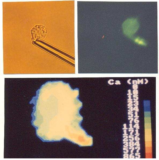
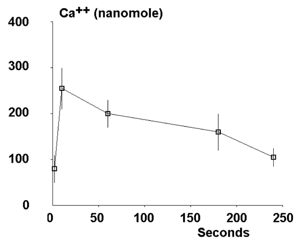
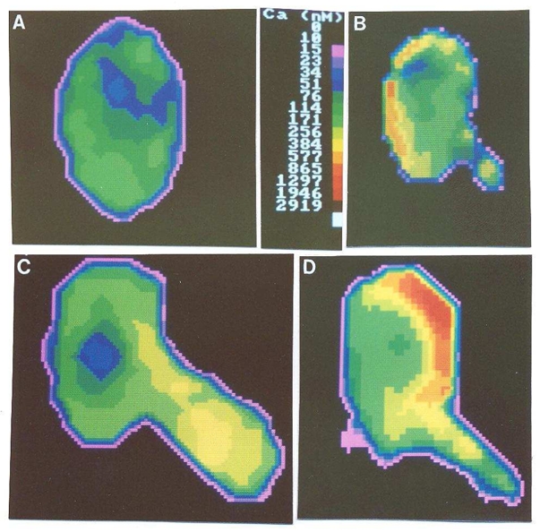
We combined fluorescence labeling, digital image processing, and micromanipulation to investigate the intracellular events induced by inflicting a mechanical stress on rat basophilic leukemia cells. Our findings were as follows: 1. Most cells displayed a localized calcium rise in response to micropipet aspiration. This represented an average threefold increase as compared to resting level, and it was observed during the first 10 s following aspiration. A slow return to initial level occurred within about 3 min. Further, this calcium rise involved a mobilization of intracellular stores, since it was not prevented by adding a calcium chelator into the extracellular medium. 2. All micropipet-aspirated cells displayed a local accumulation of microfilaments, with a preferential localization in the cell protrusions or near the pipet tips. 3. No absolute correlation was found between the localization of calcium rise and cytoskeletal accumulation. 4. Cell deformability was decreased when intracellular calcium was maintained at a constant (high or low) level with ionomycin and/or EGTA. It is concluded that cells have a general ability to respond to mechanical stimulation by a coordinated set of events. More parameters must be studied before the mechanisms of cell shape regulation are fully understood.
Figures
Similar articles
-
Role of calcium in the shape control of human granulocytes.Blood Cells. 1993;19(1):115-29; discussion 129-31. Blood Cells. 1993. PMID: 8400307
-
Release of calcium from intracellular stores in rat basophilic leukemia cells monitored with the fluorescent probe chlortetracycline.J Cell Physiol. 1990 Jan;142(1):78-88. doi: 10.1002/jcp.1041420111. J Cell Physiol. 1990. PMID: 1688862
-
Influx of extracellular calcium is required for the membrane translocation of 5-lipoxygenase and leukotriene synthesis.Biochemistry. 1991 Sep 24;30(38):9346-54. doi: 10.1021/bi00102a030. Biochemistry. 1991. PMID: 1654095
-
Changes in membrane-microfilament interaction in intercellular adherens junctions upon removal of extracellular Ca2+ ions.J Cell Biol. 1986 May;102(5):1832-42. doi: 10.1083/jcb.102.5.1832. J Cell Biol. 1986. PMID: 3084500 Free PMC article.
-
Cytosolic free Ca2+ in daunorubicin and vincristine resistant Ehrlich ascites tumor cells. Drug accumulation is independent of intracellular Ca2+ changes.Biochem Pharmacol. 1991 Jan 15;41(2):243-53. doi: 10.1016/0006-2952(91)90483-l. Biochem Pharmacol. 1991. PMID: 1899193
Cited by
-
Single cell deposition and patterning with a robotic system.PLoS One. 2010 Oct 21;5(10):e13542. doi: 10.1371/journal.pone.0013542. PLoS One. 2010. PMID: 21042403 Free PMC article.
-
How Cells feel their environment: a focus on early dynamic events.Cell Mol Bioeng. 2008 Mar;1(1):5-14. doi: 10.1007/s12195-008-0009-7. Cell Mol Bioeng. 2008. PMID: 21151920 Free PMC article.
-
Mechanically stimulated cytoskeleton rearrangement and cortical contraction in human neutrophils.Biophys J. 1995 May;68(5):2004-14. doi: 10.1016/S0006-3495(95)80377-2. Biophys J. 1995. PMID: 7612842 Free PMC article.
-
Biophysical description of multiple events contributing blood leukocyte arrest on endothelium.Front Immunol. 2013 May 15;4:108. doi: 10.3389/fimmu.2013.00108. eCollection 2013. Front Immunol. 2013. PMID: 23750158 Free PMC article.
-
Mechanotransduction as a major driver of cell behaviour: mechanisms, and relevance to cell organization and future research.Open Biol. 2021 Nov;11(11):210256. doi: 10.1098/rsob.210256. Epub 2021 Nov 10. Open Biol. 2021. PMID: 34753321 Free PMC article. Review.
References
-
- Erickson CA, Trinkaus JP. Exp Cell Res. 1976;99:375–384. - PubMed
-
- Takeuchi S. Developmental Biol. 1979;70:232–240. - PubMed
-
- de Witt MT, Handley CJ, Woakes B, Lowther DA. Connective tissue res. 1984;12:97–109. - PubMed
-
- Franke RP, Grafe M, Schnittler H, Seiffe D, Mittermayer C. Nature. 1984;307:648–649. - PubMed
Publication types
MeSH terms
Substances
LinkOut - more resources
Full Text Sources