Review
. 2006:92:225-305.
doi: 10.1016/S0065-2776(06)92006-9.

Antigen presentation and the ubiquitin-proteasome system in host-pathogen interactions

Affiliations
Review

Antigen presentation and the ubiquitin-proteasome system in host-pathogen interactions

Joana Loureiro et al. Adv Immunol. 2006.

Abstract

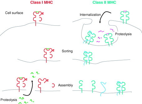
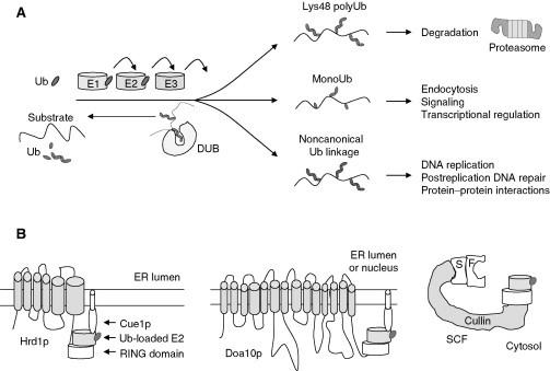
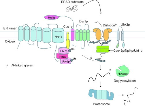
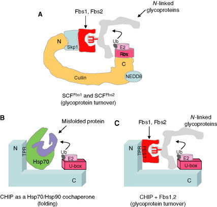
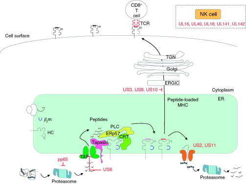
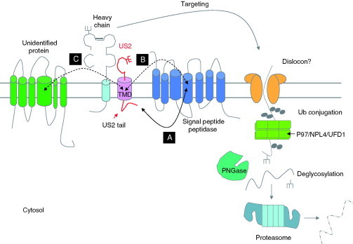
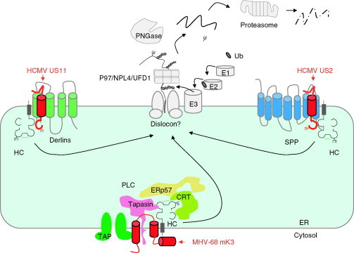
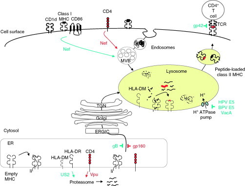
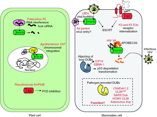
Relatively small genomes and high replication rates allow viruses and bacteria to accumulate mutations. This continuously presents the host immune system with new challenges. On the other side of the trenches, an increasingly well-adjusted host immune response, shaped by coevolutionary history, makes a pathogen's life a rather complicated endeavor. It is, therefore, no surprise that pathogens either escape detection or modulate the host immune response, often by redirecting normal cellular pathways to their advantage. For the purpose of this chapter, we focus mainly on the manipulation of the class I and class II major histocompatibility complex (MHC) antigen presentation pathways and the ubiquitin (Ub)-proteasome system by both viral and bacterial pathogens. First, we describe the general features of antigen presentation pathways and the Ub-proteasome system and then address how they are manipulated by pathogens. We discuss the many human cytomegalovirus (HCMV)-encoded immunomodulatory genes that interfere with antigen presentation (immunoevasins) and focus on the HCMV immunoevasins US2 and US11, which induce the degradation of class I MHC heavy chains by the proteasome by catalyzing their export from the endoplasmic reticulum (ER)-membrane into the cytosol, a process termed ER dislocation. US2- and US11-mediated subversion of ER dislocation ensures proteasomal degradation of class I MHC molecules and presumably allows HCMV to avoid recognition by cytotoxic T cells, whilst providing insight into general aspects of ER-associated degradation (ERAD) which is used by eukaryotic cells to purge their ER of defective proteins. We discuss the similarities and differences between the distinct pathways co-opted by US2 and US11 for dislocation and degradation of human class I MHC molecules and also a putatively distinct pathway utilized by the murine herpes virus (MHV)-68 mK3 immunoevasin for ER dislocation of murine class I MHC. We speculate on the implications of the three pathogen-exploited dislocation pathways to cellular ER quality control. Moreover, we discuss the ubiquitin (Ub)-proteasome system and its position at the core of antigen presentation as proteolysis and intracellular trafficking rely heavily on Ub-dependent processes. We add a few examples of manipulation of the Ub-proteasome system by pathogens in the context of the immune system and such diverse aspects of the host-pathogen relationship as virus budding, bacterial chromosome integration, and programmed cell death, to name a few. Finally, we speculate on newly found pathogen-encoded deubiquitinating enzymes (DUBs) and their putative roles in modulation of host-pathogen interactions.


Figures

Figure 1
Figure 1
Common principles in antigen processing and presentation by class I and class II MHC molecules. In the class I pathway, endogenous antigens are derived from cytosolic proteolysis and delivered to the ER lumen, where loading onto class I MHC products takes place. The assembled complex is then sorted to the cell surface. In the class II pathway, exogenous material is internalized from the extracellular space and delivered to the lysosome, where processing and loading onto MHC products occur. Sorting through the secretory pathway then delivers the class II complex to the cell surface.
Figure 2
Figure 2
Overview of the Ub‐proteasome system. (A) Ubiquitin‐conjugation cascade and how Ub chain linkage type and length influence substrate fate. E1, Ub‐activating enzyme; E2, Ub‐conjugating enzyme; E3, Ub‐ligase enzyme; and DUB, deubiquitinating enzyme. (B) Diversity in E3 ligases. E3s play crucial roles in substrate selection and can be regulated by localization, oligomerization, associated E2s, posttranslational modifications, and degradation. Hrd1p and Doa10p are yeast E3 ligases that are multispanning membrane proteins of the ER and, in the case of Doa10p, nuclear envelope. The mammalian SCF family of E3 ligases are mainly cytosolic and can recruit substrate adaptor proteins, the F‐boxes, with very diverse substrate specificities.
Figure 3
Figure 3
The yeast Hrd1p/Der3p‐Hrd3p E3 ligase participates in multiple steps of ERAD, from substrate selection in the ER membrane or lumen to ubiquitination in the cytosol. PNGase, peptide‐N‐glycanase; Ub, ubiquitin. See text for details.
Figure 4
Figure 4
Substrate specificity in mammalian ERAD E3s involved in glycoprotein turnover. (A) The cytosolic SCF E3 ligase catalyzes ubiquitination of dislocated N‐linked glycoproteins when complexed with the F‐box proteins Fbs1 and Fbs2; Rbx, RING‐box domain. (B) CHIP, C‐terminus of Hsc70‐interacting protein, is usually a cochaperone for the heat shock protein (Hsp) chaperone system Hsp70/Hsp90, ubiquitinating misfolded proteins bound to Hsps. (C) When complexed with Fbs1 or Fbs2, CHIP ubiquitinates dislocated glycoproteins. E2, Ub‐conjugating enzyme; Ub, ubiquitin.
Figure 5
Figure 5
HCMV interference with class I MHC antigen presentation. HCMV immunoevasins aimed at inhibition of cytotoxicity by CD8+ T cells and NK cells are in red. TCR, T cell receptor; PLC, peptide‐loading complex; TAP, transporter associated with antigen presentation; CRT, calreticulin.
Figure 6
Figure 6
The US2 dislocation pathway requires signal peptide peptidase. The US2 cytosolic tail recruits SPP (A), which is required for dislocation of class I MHC HC. An additional step critical for dislocation is dependent on interactions involving the US2 transmembrane domain (US2 TMD) and SPP (B) or other protein(s) so far unidentified (C). Ub, ubiquitin; TMD, transmembrane domain. PNGase, peptide‐N‐glycanase.
Figure 7
Figure 7
Three viral immunoevasins that co‐opt distinct ERAD pathways. The HCMV US11 immunoevasin delivers class I MHC HC molecules to Derlins for dislocation from the ER membrane and degradation, whereas HCMV US2 uses a pathway that is dependent on signal peptide peptidase. The MHV‐68 mK3 immunoevasin is an E3 ligase that uses the PLC as a platform to target murine class I MHC molecules for ubiquitination and degradation. Although the three pathways are superficially similar, the substrate selection and targeting steps at the ER membrane are very distinct. Ub, ubiquitin; E1, Ub‐activating enzyme; E2, Ub‐conjugating enzyme; E3, Ub‐ligase enzyme; PNGase, peptide‐N‐glycanase; SPP, signal peptide peptidase; TAP, transporter associated with antigen presentation; CRT, calreticulin.
Figure 8
Figure 8
Pathogen interference with class II MHC antigen presentation. TCR, T cell receptor; PLC, peptide‐loading complex. Nef, HIV‐1 Nef; gp42, EBV glycoprotein 42; BPV E5, bovine papillomavirus protein E5; HPV E5, human papillomavirus protein E5; VacA, H. pylori VacA toxin; US2, HCMV US2; Vpu, HIV‐1 Vpu; gp160, HIV‐1 glycoprotein 160; MVB, multivesicular body; Ii, invariant chain. The immunoevasins and pathways depicted in red take place in the effector CD4+ T cell, whereas those in blue occur in the antigen‐presenting cell.
Figure 9
Figure 9
Pathogen interference with the Ub‐proteasome system. Pathogens interfere with the Ub‐proteasome system not only to manipulate antigen presentation and other aspects of the immune system, but for processes as distinct as chromosomal integration, virus budding, RNA interference, and many others. They may rely on hijacking of host E3 ligase activities or encode their own. Pathogens can manipulate host DUBs as well as encode DUB activities. The function of pathogen‐encoded DUBs remains a mistery.

Similar articles

Cited by

References

    1. Adams J. The proteasome: Structure, function, and role in the cell. Cancer Treat. Rev. 2003;29(Suppl. 1):3–9. - PubMed
    1. Ahn K., Angulo A., Ghazal P., Peterson P.A., Yang Y., Fruh K. Human cytomegalovirus inhibits antigen presentation by a sequential multistep process. Proc. Natl. Acad. Sci. USA. 1996;93(20):10990–10995. - PMC - PubMed
    1. Ahn K., Gruhler A., Galocha B., Jones T.R., Wiertz E.J., Ploegh H.L., Peterson P.A., Yang Y., Fruh K. The ER‐luminal domain of the HCMV glycoprotein US6 inhibits peptide translocation by TAP. Immunity. 1997;6(5):613–621. - PubMed
    1. Aiken C., Konner J., Landau N.R., Lenburg M.E., Trono D. Nef induces CD4 endocytosis: Requirement for a critical dileucine motif in the membrane‐proximal CD4 cytoplasmic domain. Cell. 1994;76(5):853–864. - PubMed
    1. Ait‐Goughoulte M., Hourioux C., Patient R., Trassard S., Brand D., Roingeard P. Core protein cleavage by signal peptide peptidase is required for hepatitis C virus‐like particle assembly. J. Gen. Virol. 2006;87(Pt. 4):855–860. - PMC - PubMed

Publication types

MeSH terms