. 2006;34(22):6640-52.
doi: 10.1093/nar/gkl878. Epub 2006 Dec 1.

Brn-3b enhances the pro-apoptotic effects of p53 but not its induction of cell cycle arrest by cooperating in trans-activation of bax expression

Affiliations

Brn-3b enhances the pro-apoptotic effects of p53 but not its induction of cell cycle arrest by cooperating in trans-activation of bax expression

Vishwanie S Budhram-Mahadeo et al. Nucleic Acids Res. 2006.

Abstract

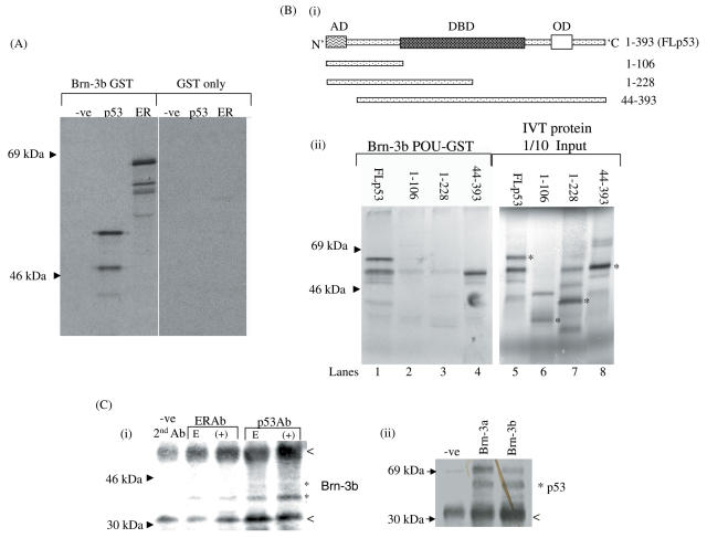
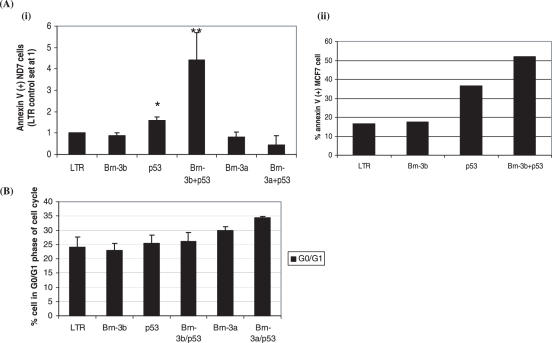
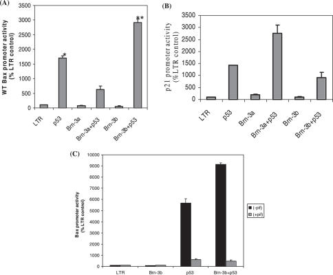
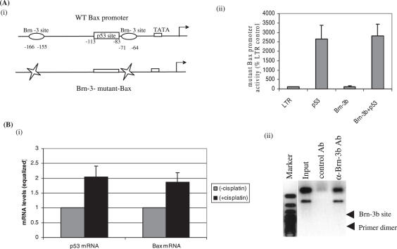
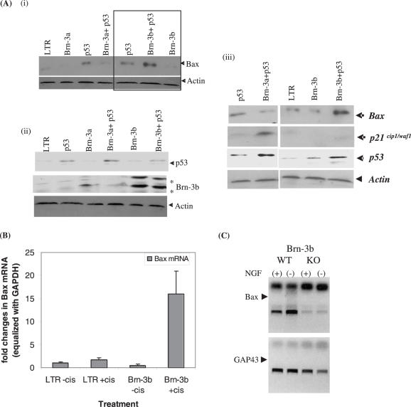
The Brn-3a and Brn-3b transcription factor have opposite and antagonistic effects in neuroblastoma cells since Brn-3a is associated with differentiation whilst Brn-3b enhances proliferation in these cells. In this study, we demonstrate that like Brn-3a, Brn-3b physically interacts with p53. However, whereas Brn-3a repressed p53 mediated Bax expression but cooperated with p53 to increase p21cip1/waf1, this study demonstrated that co-expression of Brn-3b with p53 increases trans-activation of Bax promoter but not p21cip1/waf1. Consequently co-expression of Brn-3b with p53 resulted in enhanced apoptosis, which is in contrast to the increased survival and differentiation, when Brn-3a is co-expressed with p53. For Brn-3b to cooperate with p53 on the Bax promoter, it requires binding sites that flank p53 sites on this promoter. Furthermore, neurons from Brn-3b knock-out (KO) mice were resistant to apoptosis and this correlated with reduced Bax expression upon induction of p53 in neurons lacking Brn-3b compared with controls. Thus, the ability of Brn-3b to interact with p53 and modulate Bax expression may demonstrate an important mechanism that helps to determine the fate of cells when p53 is induced.


Figures

Figure 1
Figure 1
Interaction of Brn-3b protein with p53, in vitro and in vivo. (A) Affinity chromatography (pull-down) analysis using full length Brn-3b GST fusion protein and 35S labelled IVT to show interaction of Brn-3b with p53. The p53 IVT protein was efficiently retained by Brn-3b (lane 2) showing similar affinity to positive control ER (lane 3) previously shown to interact with Brn-3b. The IVT luciferase protein (–ve control) was not retained by Brn-3b (lane 1). This interaction was specific to Brn-3b as none of the IVT proteins were retained by the GST moiety only. (B) (i) Schematic representation of the p53 deletion constructs used to identify the region of p53 required for interaction with Brn-3b POU domain. The positions of the amino acid shown represent those that are present with the deletions; AD, activation domain; DBD, DNA binding domain; OD, oligomerization domain (ii) Affinity chromatography analysis using Brn-3b POU–GST fusion protein showed that the isolated POU domain was sufficient to interact with WT FLp53 (lane 1). Use of the IVT truncated p53 constructs demonstrated that the amino acids between 1 and 106 (lane 2) or 1 and 228 (lane 3) of p53 were insufficient to mediate interaction with Brn-3b but the truncated protein, 228–393 lacking only the amino terminus (lane 4) could interact with affinity similar to the FLp53 protein. One-tenth of the input of IVT proteins used for the ‘pull-down’ assays are shown in the next panel (lanes 5–8) and * is used to indicate the size of expected polypeptide following IVT of the truncated constructs. (C) Co-immunoprecipitation assay demonstrating that Brn-3b can be isolated as a complex with p53 protein from cellular extracts. (i) IP undertaken using cellular extracts taken from neuroblastoma cells with endogenous Brn-3b (E) or with transiently transfected with Brn-3b (+), using antibodies to either ER (ER Ab) or p53 (p53 Ab) resulted in isolation of Brn-3b protein which was detected by western blotting. The control antibody (−ve) did not result in IP of Brn-3b. The isolated Brn-3b proteins are indicated by * and shows the shorter Brn-3b protein (∼32 kDa) and longer 43 kDa Brn-3b isoforms (very weakly). The non-specific bands indicated by less than appear to be associated with use of the protein A/G Sepharose to immobilize the antibody bound proteins. (ii) Immunoprecipitation of p53 with either Brn-3a or Brn-3b from cellular extracts prepared from ND7 cells transfected with Brn-3a and p53 (lane 2) or Brn-3b and p53 (lane 3). The p53 protein was detected by western blotting. The p53 protein did not immuno-precipitate with the control actin antibody (−ve) (lane 1).
Figure 2
Figure 2
Co-expression of Brn-3b with p53 increases apoptosis but did not alter cell cycle arrest in ND7 cells (A) (i) Representative changes in early apoptotic (annexin V positive), transfected (GFP positive) ND7 neuroblastoma cells as measured by FACS analysis following transfection of cells with either p53, or Brn-3b or Brn-3a alone, or p53 co-transfected with either Brn-3b or Brn-3a control. Whereas p53 alone increased percentage of apoptotic cells; this was significantly increased upon co-expression with Brn-3b compared with p53 alone (denoted by **). In contrast, co-transfection of the positive control Brn-3a with p53 leads to protection from apoptosis. Data is expressed relative to LTR control (set at 1) and represents the mean ± standard error from three independent experiments. Statistical significance was determined using Students t-test. (ii) Similar effects were seen in MCF7 cells transfected with Brn-3b or p53 either alone or together. p53 alone resulted in increased cell death compared with control transfected cells (LTR) but upon co-expression of Brn-3b with p53, the percentage of apoptotic cells was increased. (B) Analysis of changes in the percentage of transfected cells undergoing cell cycle arrest (in G0/G1) following transfection with p53 and/or Brn-3b. Brn-3a + p53 was included as the positive control. DNA content was measured by FACS analysis following propidium iodide labeling. Although p53 alone resulted in a small increase in cells accumulating in Go/G1 phase of the cell cycle, co-expression Brn-3b did not alter its effects compared with the significant changes seen upon co-expression of p53 with Brn-3a. The results represent mean (± SE) of percentage of GFP positive cells in the G0/G1 phase of the cell cycle in four independent experiments.
Figure 3
Figure 3
Co-expression of Brn-3b with p53 increases Bax promoter activity but did not alter p21cip1/waf1 promoter activity in ND7 cells (A) Effect of Brn-3b and p53 expression vectors on expression of the Bax promoter driving a luciferase gene reporter in ND7 cells. p53 strongly activated the Bax promoter (lane 2), but Brn-3a and Brn-3b mildly inhibited its basal activity (lanes 3 and 5) compared with the control vector (lane 1). As expected, Brn-3a repressed p53 mediated activity of the promoter (lane 4), but co-transfection of Brn-3b with p53 (lane 6) resulted in a significant enhancement in promoter activity compared with p53 alone (lane 2) (P < 0.05). Values are expressed as a percentage of the empty LTR control vectors. All values were equalized on the basis of the activity observed upon co-transfection with a control renilla expression vector. Values are the average of five experiments whose SD is shown by the bars. (B) Effect of Brn-3b and/or p53 expression vectors on the activity of the p21CIP1/Waf1 promoter driving a luciferase reporter gene in ND7 cells. As expected, p53 alone transactivated the promoter (lane 2) and Brn-3a mildly activated the promoter (lane 3) but cooperated with p53 to enhance the promoter activity (lane 4) compared with p53 alone only (lane 2). However, Brn-3b alone had no effect on promoter activity (lane 5) but it appears to reduce p53 mediated activation on this promoter (lane 6) compared with p53 alone. Values are expressed as a percentage of the activity obtained with the empty LTR expression vector alone (which was set at 100%). All values were equalised on the basis of the activity of a control renilla reporter plasmid which was co- transfected in all cells. Values are the average of five experiments ± SD (shown by error bars). (C) The ability of Brn-3b to increase Bax promoter activity is dependent on p53 since the p53 inhibitor, α-pifitrin, prevented the transactivation seen by p53 alone (set 3) or upon co-operation of Brn-3b and p53 (set 4). The changes in promoter activity following treatment with α-pifitrin are expressed as percentage of α-pifitrin treated LTR control (set at 100%) whereas induction in the absence of α-pifitrin is expressed as percentage of untreated LTR control (also set at 100%).
Figure 4
Figure 4
Brn-3b binds to specific sites in the Bax promoter for cooperative effects with p53 (A) (i) Schematic diagram to show the positions of the p53 binding site and the two Brn-3 binding sites within the promoter construct used that were altered in mutant Bax promoter. (ii) Reporter assays following co-transfection of Brn-3b and / or p53 with mutant Bax promoter (mutated Brn-3 sites but intact p53 binding site) into ND7 cells showed that the Brn-3 sites within the Bax promoter are required for Brn-3b to repress p53 mediated transcription of this promoter. Co-transfection of p53 resulted in transactivation of the bax promoter (lane 2) compared with control (lane 1) but Brn-3b alone had little effect (lane 3) and also failed to cooperate with p53 to further enhance promoter activity (lane 4). (B) (i) Induction of p53 and bax mRNA following treatment of ND7 cells with cisplatin as measured by qRT–PCR. Following treatment with cisplatin p53 mRNA was increased by up to 2-fold compared with untreated cells (set at 1). Similarly bax mRNA showed a corresponding increase following treatment. The results represent the mean (± SD) of three sets of treatments. (ii) Chromatin Immuno-precipitation (ChIP) of Brn-3b in cells treated with cisplatin to induce bax expression resulted in significant binding of Brn-3b to a site within the bax promoter. Amplification of a 180 bp region of the promoter which flanks the Brn-3 and p53 sites resulted in a strong band (lane 4) comparable to the input lane (lane 2). ChIP using the control actin antibody did not give rise to this band following PCR amplification (lane 3).
Figure 5
Figure 5
Co-expression of Brn-3b with p53 increases endogenous Bax expression (A) (i) Changes in the levels of Bax protein following transfection of ND7 cells with Brn-3b and p53 (lane 6) compared with p53 alone (lane 3) or Brn-3b alone (lane 7). LTR vector alone (lane 1) or Brn-3a or p53 (lanes 2 and 3) are also shown and compared with co-expression of Brn-3a and p53 (lane 4). The levels of control actin protein demonstrate protein loading in each transfected cell population. (ii) Analysis of the levels of Brn-3b and p53 to show the increase in expressed Brn-3b levels when cells were transfected with Brn-3b (lanes 5 and 6) or p53 (lanes 2, 4, 6) alone or in combination with other proteins. (iii) Analysis of bax and p21CIP1/WAF1 proteins following co-transfection of Brn-3b with p53 demonstrates that Brn-3b only cooperates with p53 to increase Bax expression (top panel, lane 5) but not and p21CIP1/WAF1(2nd panel, lane 5). This is compared with the effects of Brn-3a and p53 (lane 2) or p53 (lane 1) (carried out in the same experiment). Actin was used to demonstrate variability in loading of protein samples. (B) Changes in the expression of Bax mRNA in control or Brn-3b over-expressing MCF7 cells treated with cisplatin to induce WT p53. Lane 1 shows the bax mRNA in untreated LTR control cells was set at 1. Following treatment, there is a 2-fold increase in bax mRNA in these control cells (lane2). Levels of bax mRNA in untreated Brn-3b over-expressing cells (lane 3) was lower but this increased significantly following treatment with cisplatin (lane 4). mRNA was quantified by qRT–PCR and variability in RNA from different treatment was equalized to GAPDH. The values represent the mean and SD from three independent experiments. (C) Analysis of Bax mRNA expression in neuronal cultures obtained from DRG prepared from either WT or Brn-3b−/− (KO) mice that have been grown in the presence (+) or absence (−) of NGF (to induce apoptosis). WT cultures showed an increase in Bax mRNA following withdrawal of NGF (lane 2) compared with controls (+NGF, lane 1). DRG cultures prepared from Brn-3b KO mice showed a decrease in Bax mRNA (lane 3) and no increase even upon NGF withdrawal (lane 4). Levels of the neuronal factor, GAP43 remains unchanged suggesting that the effect is specific to bax expression.

Similar articles

Cited by

References

    1. Budhram-Mahadeo V.S., Latchman D.S. Targeting Brn-3b in breast cancer therapy. Expert Opin. Ther. Targets. 2006;10:15–25. - PubMed
    1. Theil T., Zechner U., Klett C., Adolph S., Moroy T. Chromosomal localization and sequences of the murine Brn-3 family of developmental control genes. Cytogenet. Cell Genet. 1994;66:267–271. - PubMed
    1. Lillycrop K.A., Budhram-Mahadeo V., Lakin N.D., Terrenghi G., Wood J.N., Polak J.M., Latchman D.S. A novel POU family transcription factor is closely related to Brn-3 but has a distinct expression pattern in neuronal cells. Nucleic Acids Res. 1992;20:5093–5096. - PMC - PubMed
    1. Smith M.D., Latchman D.S. The functionally antagonistic POU family transcription factors Brn-3a and Brn-3b show opposite changes in expression during the growth arrest and differentiation of human neuroblastoma cells. Int. J. Cancer. 1996;67:653–660. - PubMed
    1. Budhram-Mahadeo V., Lillycrop K.A., Latchman D.S. The levels of the antagonistic POU family transcription factors Brn-3a and Brn-3b in neuronal cells are regulated in opposite directions by serum growth factors. Neurosci. Lett. 1995;185:48–51. - PubMed

Publication types

MeSH terms

Substances