. 2007 Feb;27(4):1442-54.
doi: 10.1128/MCB.01298-06. Epub 2006 Dec 4.

RANK overexpression in transgenic mice with mouse mammary tumor virus promoter-controlled RANK increases proliferation and impairs alveolar differentiation in the mammary epithelia and disrupts lumen formation in cultured epithelial acini

Affiliations

RANK overexpression in transgenic mice with mouse mammary tumor virus promoter-controlled RANK increases proliferation and impairs alveolar differentiation in the mammary epithelia and disrupts lumen formation in cultured epithelial acini

Eva Gonzalez-Suarez et al. Mol Cell Biol. 2007 Feb.

Abstract

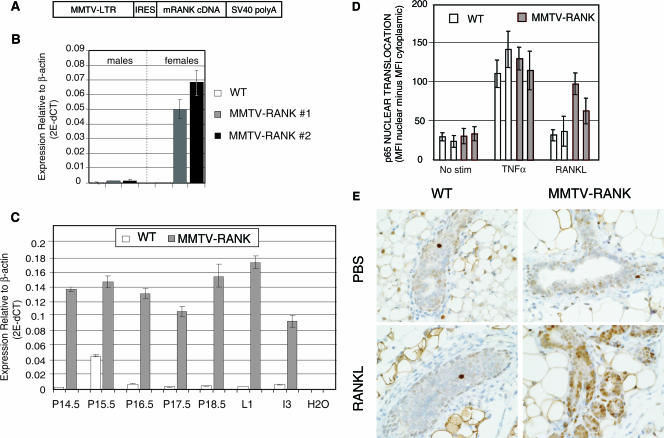
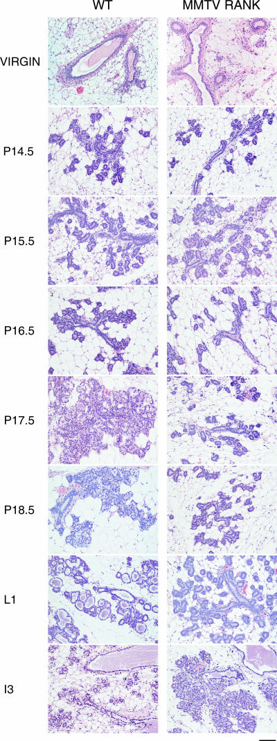
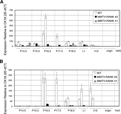
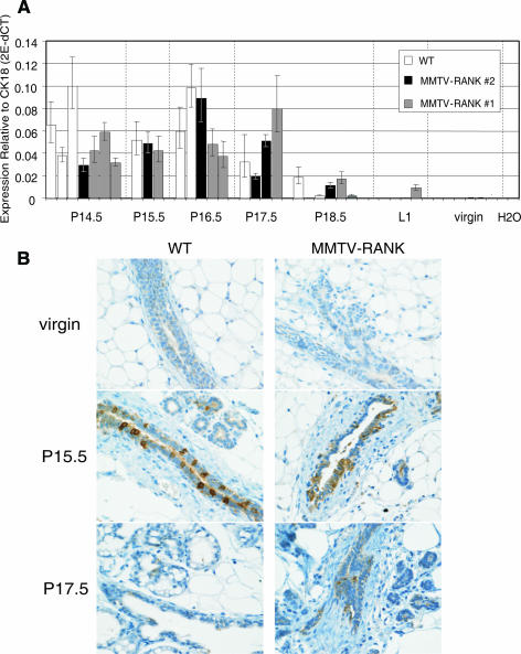
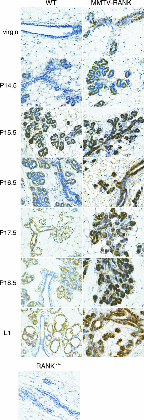
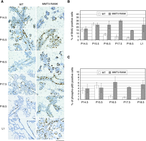
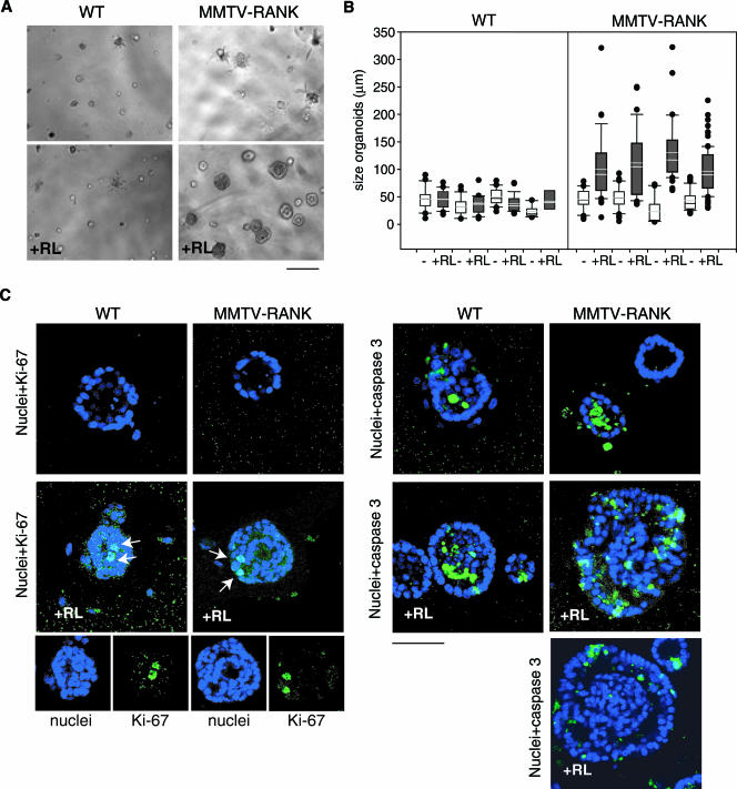
RANK and RANKL, the key regulators of osteoclast differentiation and activation, also play an important role in the control of proliferation and differentiation of mammary epithelial cells during pregnancy. Here, we show that RANK protein expression is strictly regulated in a spatial and temporal manner during mammary gland development. RANK overexpression under the control of the mouse mammary tumor virus (MMTV) promoter in a transgenic mouse model results in increased mammary epithelial cell proliferation during pregnancy, impaired differentiation of lobulo-alveolar structures, decreased expression of the milk proteins beta-casein and whey acidic protein, and deficient lactation. We also show that treatment of three-dimensional in vitro cultures of primary mammary cells from MMTV-RANK mice with RANKL results in increased proliferation and decreased apoptosis in the luminal area, resulting in bigger acini with filled lumens. Taken together, these results suggest that signaling through RANK not only promotes proliferation but also inhibits the terminal differentiation of mammary epithelial cells. Moreover, the increased proliferation and survival observed in a three-dimensional culture system suggests a role for aberrant RANK signaling during breast tumorigenesis.


Figures

FIG. 1.
FIG. 1.
Overexpression of RANK in MMTV-RANK mice and increased response to RANKL in MMTV-RANK mammary epithelial cells. (A) Schematic diagram of the MMTV-RANK transgene construct including MMTV-LTR, the rat VL30 retrotransposon internal ribosome entry site (IRES), mouse RANK CDS gene, and the SV40 polyadenylation sequences. (B) mRANK mRNA expression levels in the mammary glands of WT and MMTV-RANK (males and females) relative to β-actin in two transgenic lines generated were measured by quantitative RT-PCR. Each bar represents the average of data for 3 to 9 mice and the standard errors are indicated. We have estimated (see Materials and Methods) that MMTV-RANK mice from line 1 carry 10 to 14 copies and MMTV-RANK mice from line 2 carry 17 to 24 copies of the transgene construct. (C) mRANK expression levels in the mammary glands of WT and MMTV-RANK females relative to β-actin at different time points during pregnancy were measured by RT-PCR. Each bar represents the values for one representative mouse. Amplifications were performed in triplicate, and the standard deviation is shown. (D) Quantitative immunostaining to measure p65 nuclear translocation in primary mammary epithelial cells from WT and MMTV-RANK mice after 30 min of treatment with TNF-α (40 ng/ml) or murine RANKL (200 ng/ml). Each bar represents cells from a single mouse. Translocation is quantified using the Cellomics software as the difference between the MFI of the nuclear area minus the MFI of the cytoplasmic area. The experiment shown is representative of three experiments. (E) Phospho-p65 IHC in WT and MMTV-RANK virgin mammary glands after 30 min of stimulation with murine RANKL. No significant increase in the phospho-p65 signal is detected in luminal WT cells after RANKL stimulation. In MMTV-RANK mice a dose of 250 μg of murine RANKL results in phospho-p65 detection in most of the mammary epithelial cells. Scale bar, 100 μm.
FIG. 2.
FIG. 2.
Impaired alveolar differentiation in MMTV-RANK mice: morphology of mammary gland development in WT and MMTV-RANK transgenic mice. Glands from virgin mice and mice at different time points during pregnancy were stained with hematoxylin and eosin. Scale bar, 100 μm.
FIG. 3.
FIG. 3.
Reduction in β-casein and WAP mRNA levels in MMTV-RANK mice. β-Casein (A) and WAP (B) mRNA expression levels relative to CK18 in the mammary glands of WT and both lines of MMTV-RANK transgenic at different time points during pregnancy are shown. Each bar represents one mouse. Amplifications were performed in triplicate, and the standard deviation is shown.
FIG. 4.
FIG. 4.
RANKL expression is comparable between WT and MMTV-RANK mammary glands. (A) RANKL mRNA expression levels relative to CK18 in the mammary glands of WT and both lines of MMTV-RANK females at different time points during pregnancy measured by quantitative RT-PCR. Each bar represents one mouse. Amplifications were performed in triplicate, and the standard deviation is shown. (B) RANKL IHC in WT and MMTV-RANK mammary glands from virgin and pregnant mice at P15.5 and P17.5. Scale bar, 100 μm.
FIG. 5.
FIG. 5.
RANK protein expression is strictly regulated in the mammary gland during gestation. Images show IHC of RANK in WT and MMTV-RANK mammary glands at different time points during pregnancy. Mammary gland from a RANK-null virgin female is included as a negative control. Scale bar, 100 μm.
FIG. 6.
FIG. 6.
Sustained proliferation and p65 activation in MMTV-RANK glands at later stages of pregnancy. (A) Proliferation in the mammary glands of WT and MMTV-RANK mice at different time points during pregnancy detected by BrdU immunostaining. Note the impaired differentiation to mature alveoli in the MMTV-RANK glands. Scale bar, 100 μm. (B) Quantification of the percentage of BrdU-positive cells versus the total number of epithelial cells through pregnancy. Each bar represents the average of two to three mice, and standard errors are shown. For each mouse five fields at a magnification of ×200 were counted. (C) Quantification of the percentage of phospho-p65-positive cells versus the total number of epithelial cells throughout pregnancy is shown. For each mouse five fields at a magnification of ×200 were counted. Each bar represents one to three mice, and standard deviations or errors are shown.
FIG. 7.
FIG. 7.
RANKL directly induces proliferation and lumen repopulation in MMTV-RANK acini. (A) Phase-contrast microscopy of WT and MMTV-RANK primary MEC 3D cultures, without or with RANKL (RL) at day 4. Scale bar, 100 μm. The images are representative of five experiments. Note the significant change in morphology of the MMTV-RANK acini in the presence of RANKL. (B) Size of WT and MMTV-RANK acini after 5 days in culture in the absence (−) or presence (+) of RANKL (RL). The upper and lower bars indicate the 90th and 10th percentiles of the data. The data points greater than the 90th or less than the 10th percentiles are plotted as individual dots. For each treatment 12 to 92 spheroids were measured. (C) Confocal images at day 5 of WT and MMTV-RANK mammary acini cultured without or with RANKL (+RL) as indicated. Nuclei were stained with DRAQ5 (blue) and Ki-67 or activated caspase 3 (green). Arrows indicate Ki-67-positive cells. One or more Ki-67-positive cells was detected in 30 to 50% of the WT acini and in 100% of the MMTV-RANK acini cultured with RANKL. Representative images are shown. Scale bar, 50 μm. Note the mislocalized or sparse apoptotic cells detected in MMTV-RANK acini cultured with RANKL and the presence of a filled lumen.

Similar articles

Cited by

References

    1. Anderson, D. M., E. Maraskovsky, W. L. Billingsley, W. C. Dougall, M. E. Tometsko, E. R. Roux, M. C. Teepe, R. F. DuBose, D. Cosman, and L. Galibert. 1997. A homologue of the TNF receptor and its ligand enhance T-cell growth and dendritic-cell function. Nature 390:175-179. - PubMed
    1. Andres, A. C., M. A. van der Valk, C. A. Schonenberger, F. Fluckiger, M. LeMeur, P. Gerlinger, and B. Groner. 1988. Ha-ras and c-myc oncogene expression interferes with morphological and functional differentiation of mammary epithelial cells in single and double transgenic mice. Genes Dev. 2:1486-1495. - PubMed
    1. Arai, F., T. Miyamoto, O. Ohneda, T. Inada, T. Sudo, K. Brasel, T. Miyata, D. M. Anderson, and T. Suda. 1999. Commitment and differentiation of osteoclast precursor cells by the sequential expression of c-Fms and receptor activator of nuclear factor kappaB (RANK) receptors. J. Exp. Med. 190:1741-1754. - PMC - PubMed
    1. Barcellos-Hoff, M. H., J. Aggeler, T. G. Ram, and M. J. Bissell. 1989. Functional differentiation and alveolar morphogenesis of primary mammary cultures on reconstituted basement membrane. Development 105:223-235. - PMC - PubMed
    1. Biswas, D. K., Q. Shi, S. Baily, I. Strickland, S. Ghosh, A. B. Pardee, and J. D. Iglehart. 2004. NF-κB activation in human breast cancer specimens and its role in cell proliferation and apoptosis. Proc. Natl. Acad. Sci. USA 101:10137-10142. - PMC - PubMed

MeSH terms

LinkOut - more resources