. 2007 Feb;27(4):1334-47.
doi: 10.1128/MCB.01909-06. Epub 2006 Dec 4.

Vascular abnormalities in mice deficient for the G protein-coupled receptor GPR4 that functions as a pH sensor

Affiliations

Vascular abnormalities in mice deficient for the G protein-coupled receptor GPR4 that functions as a pH sensor

Li V Yang et al. Mol Cell Biol. 2007 Feb.

Abstract

GPR4 is a G protein-coupled receptor expressed in the vasculature, lung, kidney, and other tissues. In vitro ectopic overexpression studies implicated GPR4 in sensing extracellular pH changes leading to cyclic AMP (cAMP) production. To investigate its biological roles in vivo, we generated GPR4-deficient mice by homologous recombination. Whereas GPR4-null adult mice appeared phenotypically normal, neonates showed a higher frequency of perinatal mortality. The average litter size from GPR4(-/-) intercrosses was approximately 30% smaller than that from GPR4(+/+) intercrosses on N3 and N5 C57BL/6 genetic backgrounds. A fraction of knockout embryos and neonates had spontaneous hemorrhages, dilated and tortuous subcutaneous blood vessels, and defective vascular smooth muscle cell coverage. Mesangial cells in kidney glomeruli were also significantly reduced in GPR4-null neonates. Some neonates exhibited respiratory distress with airway lining cell metaplasia. To examine whether GPR4 is functionally involved in vascular pH sensing, an ex vivo aortic ring assay was used under defined pH conditions. Compared to wild-type aortas, microvessel outgrowth from GPR4-null aortas was less inhibited by acidic extracellular pH. Treatment with an analog of cAMP, a downstream effector of GPR4, abolished microvessel outgrowth bypassing the GPR4-knockout phenotype. These results suggest that GPR4 deficiency leads to partially penetrant vascular abnormalities during development and that this receptor functions in blood vessel pH sensing.


Figures

FIG. 1.
FIG. 1.
Generation of GPR4-knockout mice. (A) Strategy for homologous recombination at the GPR4 gene locus. The coding region of GPR4 was replaced by the IRES-GFP and PGK-neo cassette. (B) Southern blot analysis of XbaI-digested genomic DNA from recombined ES clones. (C) PCR genotyping of tail genomic DNA from mice generated by heterozygote (Het) crosses. (D) RT-PCR assay of RNA from GPR4−/− (knockout) mice and the GPR4+/+ (wild-type) littermate control. RNA was extracted from the lung, kidney, and heart and subjected to RT-PCR with gene-specific primers. KO, knockout; WT, wild type.
FIG. 2.
FIG. 2.
Litter sizes of GPR4 mouse crosses. Pup numbers delivered were counted on the first day after birth. (A) Litter sizes of different genotype crosses on the N3 backcrossed C57BL/6 genetic background. (B) Litter sizes of GPR4+/+ (wild-type) and GPR4−/− (knockout) intercrosses on the N5 backcrossed C57BL/6 genetic background. Each dot represents one litter. WT, wild type; KO, knockout; F, female; M, male.
FIG. 3.
FIG. 3.
Macroscopic hemorrhaging in GPR4-null embryos and neonates. (A to F) Mouse embryos at different developmental stages. Note hemorrhages (arrows) in the head and limb and under the skin in a fraction of GPR4−/− embryos but not GPR4+/+ embryos. (G to P) Some GPR4−/− neonates died perinatally. Internal bleeding; hemorrhages in the lung, kidney, or skin; and abnormal mesenteric blood vessels were observed (arrows). P1, postnatal day 1. Bars, 2 mm.
FIG. 4.
FIG. 4.
Histological analysis of GPR4-null embryos and neonates. (A and B) Hematoxylin and eosin staining was performed for the head tissue sections of the 11.5-dpc embryos. In the GPR4+/+ embryo, fetal red blood cells were well contained in the blood vessel (A, arrow). In the GPR4−/− embryo with cerebral hemorrhaging, blood cells were leaked into the mesenchyme (B, arrows). Magnification, ×200. (C and D) Hematoxylin and eosin staining of neonatal lungs. Hemorrhages were observed in the GPR4−/− lung (D, arrow), and sacculi were poorly expanded. Magnification, ×100. (E and F) Hematoxylin and eosin staining of neonatal kidneys. Hemorrhages were detected in the glomeruli and parenchyma of GPR4-null neonatal kidneys (F, arrows). gl, glomerulus. Magnification, ×200. Bars, 100 μm.
FIG. 5.
FIG. 5.
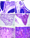
Lung anomalies in GPR4-null neonates. (A) Some GPR4-null neonates showed respiratory distress, turned cyanotic, and died on the first day (P1) after birth. (B to G) Hematoxylin and eosin staining of GPR4+/+ and GPR4−/− mouse neonatal lung paraffin sections. The wild-type bronchioles (arrows) were lined with a single layer of epithelial cells with normal morphology (B and C). In contrast, the knockout bronchioles (arrows) were lined with metaplastic clear-cytoplasm epithelial cells (arrowhead), and some regions had multiple layers (D and E). In the neonate with no obvious respiratory stress, the phenotype of epithelial cell metaplasia in bronchioles was less severe (F and G). (H and I) Hematoxylin and eosin staining of GPR4+/+ and GPR4−/− adult lung paraffin sections. No clear-cytoplasm epithelial cells were detected in adult knockout bronchioles (arrows). Magnifications, ×200 (B, D, F, H, and I) and ×400 (C, E, and G). WT, wild type; KO, knockout. Bars, 50 μm.
FIG. 6.
FIG. 6.
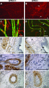
Blood vessel abnormalities and defective smooth muscle coverage in GPR4-null mice. (A and B) Whole-mount CD31 staining was performed with skins from GPR4+/+ and GPR4−/− neonates. Compared to the homogenous and hierarchical vasculature in the wild-type skin (A), tortuous and dilated blood vessels were observed in GPR4-null neonates (B, arrows). Magnification, ×100. (C and D) Blood vessels in the wild-type and GPR4-null skins were double stained with CD31 (red) and αSMA (green). Dilated blood vessels and weaker smooth muscle cell coverage (D, arrows) were evident in GPR4-null mice. Magnification, ×400. (E to H) Paraffin sections of mouse embryos were stained for αSMA as brown signals and counterstained with hematoxylin. Compared to the continuous smooth muscle cell coverage of blood vessels in the wild-type embryo (E, arrow), some GPR4−/− blood vessels were poorly coated with smooth muscle cells (F, arrows). Note the ruptured vessel with few associated αSMA-positive cells (H, arrow). de, dermis, sk, skeletal muscle. Magnifications, ×400 (E and F) and ×1,000 (G and H). (I and J) Smooth muscle cell coverage of larger arteries was similar in wild-type and knockout embryos. Magnification, ×400. Bars, 50 μm.
FIG. 7.
FIG. 7.
Mesangial cell defects in GPR4-null neonatal glomeruli. Sections of neonatal kidneys (postnatal day 1) were stained with αSMA antibodies. (A to D) Paraffin sections were immunostained for αSMA as brown signals and counterstained with hematoxylin. Compared to wild-type glomeruli (A and C, arrows), αSMA-positive mesangial cells were significantly reduced in knockout glomeruli (B and D, arrows), whereas the αSMA staining of small arteries and arterioles (arrowheads) was similar. Peritubular interstitial cells were also decreased in knockout kidneys (blue arrowheads). Magnifications, ×400 (A and B) and ×1,000 (C and D). (E and F) Vibratome sections of neonatal kidneys were double stained with CD31 (red) and αSMA (green). αSMA-positive mesangial cells were diminished in GPR4-null glomeruli (F, arrows), but the smooth muscle cell coverage of small arteries (arrowheads) appeared normal. Endothelial compartments in wild-type and knockout glomeruli were similar. Magnification, ×400. Bars, 25 μm. gl, glomerulus; ar, artery; rt, renal tube.
FIG. 8.
FIG. 8.
Microvessel outgrowth from GPR4−/− aortic rings is less responsive to the inhibitory effect of acidic pH. (A) Aortic rings were embedded in Matrigel and cultured for 4 days in EGM-2 medium with different pHs or with 500 μM 8-bromo-cAMP. Outgrowth of microvessels was examined under an invert microscope at a magnification of ×40. Similar patterns of pH effects were observed in three independent experiments. (B) Quantification of microvessel outgrowth. The outgrowth area was encircled and quantified with the ImageJ software as described in Materials and Methods. Data were presented as means ± standard errors of the means from three to five aortic rings. The outgrowth of GPR4+/+ aortic rings at pH 7.5 was set as the control (100%).

References

    1. An, S., C. Tsai, and E. J. Goetzl. 1995. Cloning, sequencing and tissue distribution of two related G protein-coupled receptor candidates expressed prominently in human lung tissue. FEBS Lett. 375:121-124. - PubMed
    1. Berger, A. C., X. Q. Wang, A. Zalatoris, J. Cenna, and J. C. Watson. 2004. A murine model of ex vivo angiogenesis using aortic disks grown in fibrin clot. Microvasc. Res. 68:179-187. - PubMed
    1. Betsholtz, C., P. Lindblom, M. Bjarnegard, M. Enge, H. Gerhardt, and P. Lindahl. 2004. Role of platelet-derived growth factor in mesangium development and vasculopathies: lessons from platelet-derived growth factor and platelet-derived growth factor receptor mutations in mice. Curr. Opin. Nephrol Hypertens. 13:45-52. - PubMed
    1. Burbridge, M. F., D. C. West, G. Atassi, and G. C. Tucker. 1999. The effect of extracellular pH on angiogenesis in vitro. Angiogenesis 3:281-288. - PubMed
    1. Carey, A. V., R. M. Carey, and R. A. Gomez. 1992. Expression of alpha-smooth muscle actin in the developing kidney vasculature. Hypertension 19(2 Suppl.):II168-II175. - PubMed

Publication types

MeSH terms

Substances