. 2006 Dec 12;103(50):19123-7.
doi: 10.1073/pnas.0607614103. Epub 2006 Dec 4.

Influenza virus pleiomorphy characterized by cryoelectron tomography

Affiliations

Influenza virus pleiomorphy characterized by cryoelectron tomography

Audray Harris et al. Proc Natl Acad Sci U S A. .

Abstract

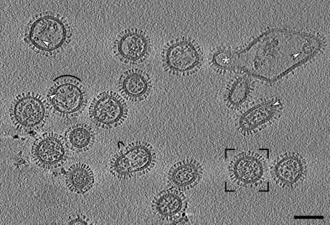
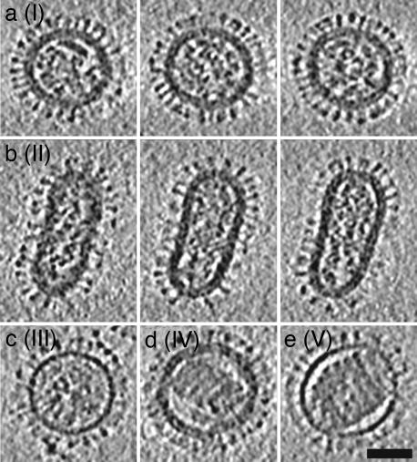
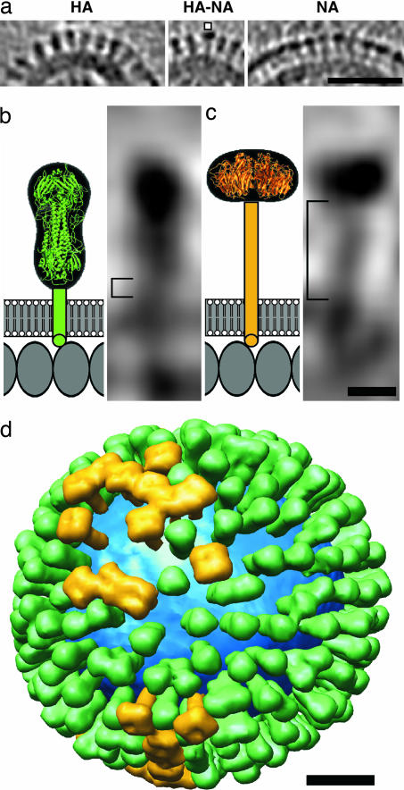
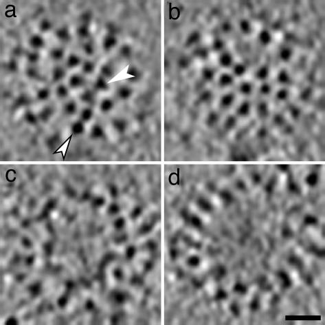
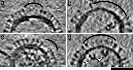
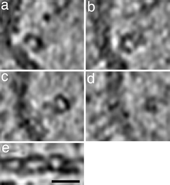
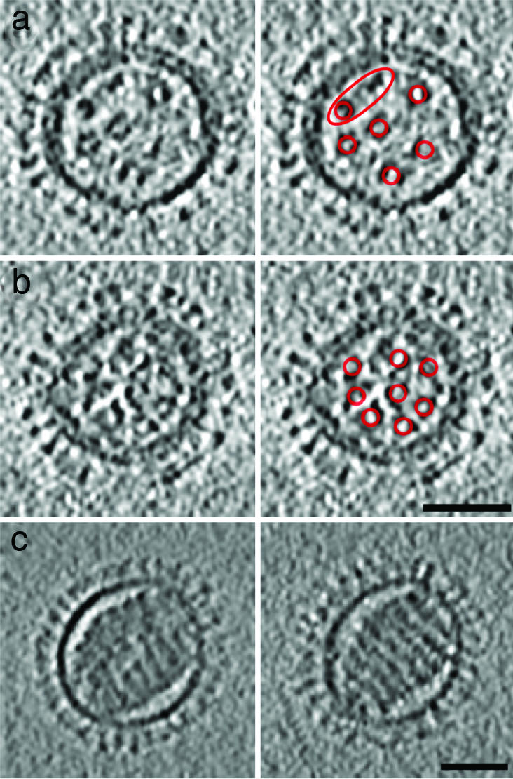
Influenza virus remains a global health threat, with millions of infections annually and the impending threat that a strain of avian influenza may develop into a human pandemic. Despite its importance as a pathogen, little is known about the virus structure, in part because of its intrinsic structural variability (pleiomorphy): the primary distinction is between spherical and elongated particles, but both vary in size. Pleiomorphy has thwarted structural analysis by image reconstruction of electron micrographs based on averaging many identical particles. In this study, we used cryoelectron tomography to visualize the 3D structures of 110 individual virions of the X-31 (H3N2) strain of influenza A. The tomograms distinguish two kinds of glycoprotein spikes [hemagglutinin (HA) and neuraminidase (NA)] in the viral envelope, resolve the matrix protein layer lining the envelope, and depict internal configurations of ribonucleoprotein (RNP) complexes. They also reveal the stems that link the glycoprotein ectodomains to the membrane and interactions among the glycoproteins, the matrix, and the RNPs that presumably control the budding of nascent virions from host cells. Five classes of virions, four spherical and one elongated, are distinguished by features of their matrix layer and RNP organization. Some virions have substantial gaps in their matrix layer ("molecular fontanels"), and others appear to lack a matrix layer entirely, suggesting the existence of an alternative budding pathway in which matrix protein is minimally involved.


Conflict of interest statement

The authors declare no conflict of interest.

Figures

Fig. 1.
Fig. 1.
Section through a cryotomogram of a field of influenza virions. Three planes were averaged to reduce noise, giving a section 4.7 nm thick. White arrowheads mark typical RNPs: at left, transverse C-shaped sections; at right, longitudinal sections through relatively straight as well as curved RNPs. Black arcs mark areas with matrix layer gaps and missing or lower density packings of glycoprotein spikes. The framed virion is shown in greater detail in Fig. 2a. The large irregular particle at top right was probably generated during cell disruption; it contains at least one budding virus (white asterisk). (Scale bar, 100 nm.)
Fig. 2.
Fig. 2.
Examples of the five morphological classes (I–V) of influenza virions. For the more abundant class I and II particles, three serial slices, 10.9 nm apart and 4.7 nm thick, are shown in a and b, respectively, whereas single near-central slices are shown for classes III-V in ce. The class I virion (a) is the framed particle in Fig. 1. (Scale bar, 50 nm.)
Fig. 3.
Fig. 3.
Distributions and shape-based differentiation of HA and NA spikes. (a) HA cluster (Left); single NA (marked) in a cluster of HAs (Center); and cluster of mainly NA spikes (Right). (Scale bar, 50 nm.) (b and c) The stem lengths of HA and NA (square brackets in b and c, respectively) were measured as described in Materials and Methods. The structures of the stems, transmembrane segments, and endodomain tails are not known, and they are shown schematically. Molecules in the matrix layer are inferred to be packed in a monolayer with a spacing of ≈4 nm (our unpublished results). (Scale bar, 5 nm.) (d) Model of the distribution of glycoprotein HA (green) and NA (gold) on a single influenza virion. The lipid bilayer is blue. (Scale bar, 20 nm.)
Fig. 4.
Fig. 4.
Patches of glycoprotein spikes depicted in tangential sections (4.7 nm thick) through virions in cryotomograms. In most cases, triangular HA spikes (e.g., white arrowhead in a) are distinguishable from square NA spikes (e.g., white arrowhead with black border in a). Spikes are close-packed, without lateral order, apart from occasional bare patches of variable size. Some examples of bare patches are shown in b–d. (Scale bar, 20 nm.)
Fig. 5.
Fig. 5.
Influenza virus particles with gaps in their matrix layer. Virions containing matrix gaps tend to have a decreased density of glycoproteins in the envelope regions overlying the gaps, which are indexed with arcs. The white arrowheads in d points to C-shaped transverse sections through two RNPs. For each particle, a 4.7-nm-thick slice is shown. (Scale bar, 50 nm.)
Fig. 6.
Fig. 6.
Distributions of density perpendicular to and along influenza virus ribonucleoprotein particles are illustrated in tomographic slices. (ad) Four serial slices, 4.7 nm thick and 9.4 nm apart, through part of an influenza virion. An RNP segment is oriented approximately perpendicular to the viewing direction. (e) A central longitudinal section of the same RNP complex, after computational straightening, suggests a double helical structure. (Scale bar, 20 nm.)
Fig. 7.
Fig. 7.
Central slices through influenza virions containing (7 + 1)-like and solenoid RNP configurations. a and b show two examples of (7 + 1)-like configurations in spherical class I virions. In each case, a slice is shown in Left and, on Right, it is indexed with red rings around the RNPs, which are mostly C-shaped in cross-section. In virion (a), which has an incomplete matrix layer, only six peripheral RNPs are clearly visible (two are enclosed in the red ellipse). Weaker density may denote a seventh peripheral RNP. (c) Two serial slices through the solenoidal core of a class V virion, which has no discrenible matrix layer. The slices are 18.7 nm apart and 4.7 nm thick. (Scale bars, 50 nm.)

References

    1. Lamb RA, Krug RM. In: Fields Virology. Knipe DM, Howley PM, editors. Vol 1. New York: Lippincott Williams and Wilkins; 2001. pp. 1487–1579.
    1. Wilson IA, Skehel JJ, Wiley DC. Nature. 1981;289:366–373. - PubMed
    1. Varghese JN, Laver WG, Colman PM. Nature. 1983;303:35–40. - PubMed
    1. Harris A, Forouhar F, Qiu S, Sha B, Luo M. Virology. 2001;289:34–44. - PubMed
    1. Schulze IT. Virology. 1972;47:181–196. - PubMed

Publication types

MeSH terms