

Is estradiol mandatory for an adequate follicular and embryo development? A mouse model using aromatase inhibitor (anastrozole)
- PMID: 17146736
- PMCID: PMC3455099
- DOI: 10.1007/s10815-006-9089-2
Is estradiol mandatory for an adequate follicular and embryo development? A mouse model using aromatase inhibitor (anastrozole)
Abstract
Background: Although high levels of estradiol are found in the follicular fluid, little is known about its necessity for adequate follicular growth, oocyte maturation and embryo development. Arimidex (anastrozole) is a potent aromatase inhibitor capable to induce an in-vivo milieu deprived of estradiol. This study uses a mouse model applying Arimidex to create an in-vivo system lacking of estradiol, in order to explore whether this gonadal steroid hormone is mandatory for folliculogenesis followed by normal fertilization and embryo development.
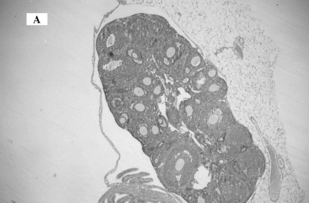
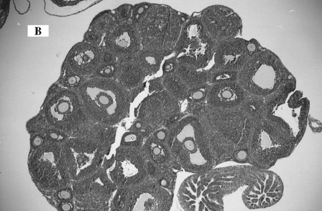
Methods: Experiment 1: Immature C57 Black female mice, aged 3-4 weeks were superovulated by 5 IU PMSG given intraperitoneally. A study group (9 mice) was concomitantly injected with 0.1 mg of Arimidex intraperitoneally given the morning day before PMSG, the morning day of PMSG injection and the following two days. The control group (8 mice) was similarly injected with normal saline. Estradiol (E2) and progesterone (P) serum levels were tested 48 hours after PMSG and the ovaries of each mouse blindly examined by a pathologist to evaluate follicular development. Experiment 2: 48 h after PMSG superovulation, hCG (7.5 IU) was injected intraperitoneally, followed by mating. The study group was treated with Arimidex 0.1 mg intraperitoneally daily from a day prior to PMSG injection to the day of sacrifice. The control group was treated similarly by normal saline. Forty-two hours after mating blood was withdrawn for E2 and P levels followed by tubal dissection. Embryos of 2-4 cells were cultured in-vitro and the development to the morula, blastocyst and hatching blastocyst stages were examined 24, 42, and 48 h later.
Results: Experiment 1: A significant reduction of E2 levels was achieved in the Arimidex group in comparison to control group (126.3+/-104.8 and 1910+/-960 pmol/L, respectively; p < 0.0001). Nevertheless, the two groups did not differ by the mean number of follicles (27+/-9.5 and 30.4+/-13.0) or the distribution for antral (65% and 68.4%) and pre-antral (35% and 31.6%) follicles, respectively. Experiment 2: The reduction of estradiol during follicular phase did not hamper follicular development, in-vivo fertilization and in-vitro embryo development. Similar rates of embryo development to the morula stage (90.6% and 86%), blastocyst stage (86% and 89%) and hatching blastocyst (81% and 78%) were achieved in the Arimidex group and the control group, respectively.
Conclusions: Adequate folliculogenesis is independent of estrogen but is conditioned on gonadotropin stimulation. Moreover, depletion of estradiol in the vicinity of the oocyte did not impair its developmental potential, including its fertilization and development into morulae, blastocysts and hatching blastocysts.
Figures
Similar articles
-
Effect of estrogen deprivation on follicle/oocyte maturation and embryo development in mice.Chin Med J (Engl). 2004 Apr;117(4):498-502. Chin Med J (Engl). 2004. PMID: 15109437
-
Effects of aromatase inhibition on in vitro follicle and oocyte development analyzed by early preantral mouse follicle culture.Mol Reprod Dev. 2002 Apr;61(4):549-59. doi: 10.1002/mrd.10107. Mol Reprod Dev. 2002. PMID: 11891927
-
Preimplantation embryo development and serum steroid levels in immature rats induced to ovulate or superovulate with pregnant mares' serum gonadotropin injection or follicle-stimulating hormone infusions.Gamete Res. 1989 May;23(1):127-38. doi: 10.1002/mrd.1120230112. Gamete Res. 1989. PMID: 2501205
-
ARIMIDEX: a new oral, once-a-day aromatase inhibitor.J Steroid Biochem Mol Biol. 1995 Jun;53(1-6):175-9. doi: 10.1016/0960-0760(95)00045-2. J Steroid Biochem Mol Biol. 1995. PMID: 7626450 Review.
-
Estrogen-regulated synaptogenesis in the hippocampus: sexual dimorphism in vivo but not in vitro.J Steroid Biochem Mol Biol. 2012 Aug;131(1-2):24-9. doi: 10.1016/j.jsbmb.2011.11.010. Epub 2011 Nov 25. J Steroid Biochem Mol Biol. 2012. PMID: 22138012 Review.
Cited by
-
Reducing 3D Hydrogel Stiffness, Addition of Oestradiol in a Physiological Concentration and Increasing FSH Concentration Improve In Vitro Growth of Murine Preantral Follicles.Int J Mol Sci. 2023 Aug 6;24(15):12499. doi: 10.3390/ijms241512499. Int J Mol Sci. 2023. PMID: 37569872 Free PMC article.
-
Synthesis and biological evaluation of two agents for imaging estrogen receptor β by positron emission tomography: challenges in PET imaging of a low abundance target.Nucl Med Biol. 2012 Nov;39(8):1105-16. doi: 10.1016/j.nucmedbio.2012.05.011. Epub 2012 Jun 30. Nucl Med Biol. 2012. PMID: 22749433 Free PMC article.
-
Hypothyroidism Reduces the Size of Ovarian Follicles and Promotes Hypertrophy of Periovarian Fat with Infiltration of Macrophages in Adult Rabbits.Biomed Res Int. 2017;2017:3795950. doi: 10.1155/2017/3795950. Epub 2017 Jan 4. Biomed Res Int. 2017. PMID: 28133606 Free PMC article.
-
A novel approach for long-term oral drug administration in animal research.J Neurosci Methods. 2011 Feb 15;195(2):194-9. doi: 10.1016/j.jneumeth.2010.12.009. Epub 2010 Dec 14. J Neurosci Methods. 2011. PMID: 21163304 Free PMC article.
-
Potential use of Indonesian basil (Ocimum basilicum) maceration to increase estradiol and progesterone synthesis and secretion to improve prenatal growth of offspring using female albino rats as an animal model.Vet World. 2022 May;15(5):1197-1207. doi: 10.14202/vetworld.2022.1197-1207. Epub 2022 May 18. Vet World. 2022. PMID: 35765474 Free PMC article.
References
-
- Hillier SG. Ovarian manipulation with pure gonadotropins. J Endocrinol. 1990;127:1–4. - PubMed
-
- Wu TC, Wang L, Wan YJ. Detection of estrogen receptor messenger ribonucleic acid in human oocytes and cumulus-oocyte complexes using reverse transcriptase-polymerase chain reaction. Fertil Steril. 1993;59:54–59. - PubMed
-
- Couse JF, Lindzey, Grandrian K, Gustafsson JA, Korach KS. Tissue distribution and quanitative analysis of estrogen receptor-α (ERα) and estrogen receptor-β (ERβ) messenger ribonucleic acid in the wild wild type and ER-α knockout mouse. Endocrinollogy 1997;138:4613–21. - PubMed
MeSH terms
Substances
LinkOut - more resources
Full Text Sources
Other Literature Sources
