. 2007 Apr 1;403(1):49-57.
doi: 10.1042/BJ20061257.

Serglycin proteoglycan is required for secretory granule integrity in mucosal mast cells

Affiliations

Serglycin proteoglycan is required for secretory granule integrity in mucosal mast cells

Tiago Braga et al. Biochem J. .

Abstract

SG (serglycin) PGs (proteoglycans) are strongly implicated in the assembly of MC (mast cell) granules. However, this notion has mainly been on the basis of studies of MCs of the connective tissue subtype, whereas the role of SG PG in mucosal MCs has not been explored. In the present study, we have addressed the latter issue by using mice with an inactivated SG gene. Bone marrow cells were differentiated in vitro into the mucosal MC phenotype, expressing the markers mMCP (mouse MC protease) -1 and -2. Biosynthetic labelling experiments performed on these cells revealed an approximately 80% reduction of 35SO4(2-) incorporation into PGs recovered from SG-/- cells as compared with SG+/+ counterparts, indicating that SG is the dominating cell-associated PG of mucosal MCs. Moreover, the absence of SG led to defective metachromatic staining of mucosal MCs, both in vivo and in the in vitro-derived mucosal MCs. Ultrastructural analysis showed that granules were present in similar numbers in SG+/+ and SG-/- cells, but that their morphology was markedly affected by the absence of SG, e.g. with electron-dense core formation only seen in SG+/+ granules. Analysis of the MC-specific proteases showed that mMCP-1 and mMCP-7 were completely independent of SG for storage, whereas mMCP-2 showed a partial dependence. In contrast, mMCP-4 and -6, and carboxypeptidase A were strongly dependent on SG for storage. Together, our data indicate that SG PG is of crucial importance for assembly of mature mucosal MC granules, but that the specific dependence on SG for storage varies between individual granule constituents.


Figures

Figure 1
Figure 1. Morphology and phenotype of mucosal-type BMMCs
(A) Cytospin slides were prepared from bone marrow cells recovered from SG+/+ and SG−/− mice after 15 days of culture in medium containing TGF-β/IL-3/IL-9/SCF. Slides were stained with May–Grünwald–Giemsa. Note that SG−/− cells do not show metachromatic staining of their granules, and note also the May–Grünwald–Giemsa-negative vesicles (arrows) present in their cytoplasm, while SG+/+ cells display metachromatically stained granules. (B) Sections were prepared from small intestines of SG+/+ and SG−/− animals and were stained with Toluidine Blue. Rare Toluidine Blue-positive cells were detected in the mucosal region (left-hand panel; arrow) and in the submucosa (right-hand panel; arrow) of SG+/+ mice. No Toluidine Blue-positive cells were found in sections prepared from SG−/− animals (results not shown). (C) Chloroacetate esterase activity in SG+/+ (left-hand panel) and SG−/− (right-hand panel) in vitro-derived MMC-like cells. Note that cells of both the SG+/+ and the SG−/− genotypes show strong chloroacetate esterase activity.
Figure 2
Figure 2. TEM of SG+/+ and SG−/− cells showing granules
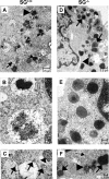
(A) Representative micrograph from SG+/+ in vitro-derived MMC-like cells demonstrating granules containing (i) many small electron dense vesicles (arrow), (ii) small vesicles together with grainy substance (arrowhead) or (iii) mainly grainy substance (broken arrow). Original magnification ×20000; scale bar, 0.5 μm. (B) High magnification clearly revealed the grainy structure and the distinction of electron-lucent/electron-dense parts of the granules in the SG+/+ cells. Original magnification ×75000; scale bar, 0.1 μm. (C) A closer view of the coexistence in SG+/+ cells of granules filled with small vesicles (arrows) and granules with an electron-dense core of a grainy material (arrowhead). Original magnification ×42000; scale bar, 0.1 μm. (D) A representative micrograph of a SG−/− cell shows cytoplasm granules (arrows), which are mostly spherical, but some are ovoid. Most of the SG−/− granules were completely filled with an amorphous matrix of moderate electron density, although solitary granules contained small vesicles harbouring material of varying electron density (arrowhead). Original magnification ×18000; scale bar, 0.5 μm. (E) The amorphous matrix of the SG−/− granules is obvious at high magnification. Original magnification ×75000; scale bar, 0.1 μm. (F) Detail of the cytoplasm co-localization of the typical SG−/− cell granules with amorphous granule matrix (arrows) and granules containing small vesicles filled with material of different electron density (arrowhead). Note the incorporation/budding of a small vesicle (broken arrow). Original magnification ×42000; scale bar, 0.1 μm.
Figure 3
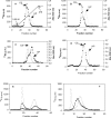
Figure 3. Analysis and characterization of 35S-labelled GAGs
SG+/+ and SG−/− BMMCs were biosynthetically labelled with 35SO42−, followed by isolation of 35S-labelled GAGs from the cell layer and from conditioned media. Samples (10000 c.p.m.) from the cell fractions of SG+/+ (A; ■) and SG−/− (B; ■) in vitro-derived MMC-like cells, and from the conditioned medium of SG+/+ cells (D; ■) were analysed by anion-exchange chromatography, and the elution positions of the 35S-labelled macromolecules were compared with those of internal standards of unlabelled CS and pig mucosal heparin (HP; CS and HP were detected by the carbazole reaction; solid grey line). Note the co-elution of GAGs from in vitro-derived MMC-like cells with standard CS. (C) GAGs isolated from SG+/+ (■) and SG−/− (◇) bone marrow cells (cell layer) that had been cultured under conditions that result in MCs of a CTMC-like phenotype [30] were analysed by anion-exchange chromatography along with internal standards of CS and heparin. Note the co-elution of GAGs from SG+/+ cells with standard heparin and the markedly lower charge density of 35S-labelled macromolecules from SG−/− cells. (E) 35S-labelled GAGs recovered from SG+/+ (□) or SG−/− (♦) in vitro-derived MMC-like cells were analysed on a Sephadex G-50 column, either before (solid line) or after (broken line) digestion with chondroitinase ABC. (F) Purified 35S-labelled macromolecules recovered from the cell layer of SG+/+ (□; solid line) or SG−/− cells (▲; solid line) were analysed by gel filtration on a Superose 6 column eluted with 0.2 M NH4HCO3. Samples of SG+/+ (□; dashed line) and SG−/− (♦; broken line) were also analysed after release of free GAG chains from the PGs by treatment with 0.5 M NaOH.
Figure 4
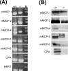
Figure 4. mRNA and protein levels of MC proteases in in vitro-derived MMC-like SG+/+ and SG−/− cells
(A) mRNA expression of MC proteases. Total RNA was isolated from SG+/+ and SG−/− bone marrow cells after 15 days of culture in medium containing TGF-β/IL-3/IL-9/SCF, and was analysed by RT–PCR, using the primers specified in the Experimental section. Expression of the HPRT gene was used as housekeeping control. Lane M, molecular-mass markers. (B) Immunoblot analysis of MC proteases in extracts from in vitro-derived MMC-like cells. SG+/+ and SG−/− BMMCs were cultured for 15 days in medium containing TGF-β/IL-3/IL-9/SCF. Cellular extracts were prepared and were analysed for the presence of the proteases indicated.
Figure 5
Figure 5. Immunoblot analysis of MC proteases in intestinal extracts
Intestinal extracts were prepared from SG+/+ (+/+) and SG−/− (−/−) mice by a two-step procedure, first with a low-salt (LS) buffer and subsequently with a high-salt (HS) buffer. Low- and high-salt extracts were analysed for the presence of the indicated proteases.
Figure 6
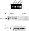
Figure 6. mRNA and immunoblot analysis of mMCP-7
(A) In vitro-derived MMC-like cells were cultured from SG+/+ and SG−/− mice of the DBA-1 genetic background. After 4 days of culture, total RNA was isolated and was analysed for mMCP-7 and HPRT (housekeeping control) mRNA expression by RT–PCR. (B) In vitro-derived MMC-like cells (taken after 15 days of culture) from SG+/+ and SG−/− mice of the DBA-1 background were analysed for mMCP-7 antigen by immunoblot analysis. As a negative control, in vitro-derived MMC-like cells derived from C57BL/6 animals were also analysed for mMCP-7 antigen. (C) Ear pinnae were prepared from SG+/+ and SG−/− mice of the DBA-1 background and were extracted with a high-salt buffer, followed by immunoblot analysis for mMCP-7 antigen.

References

    1. Ha T. Y., Reed N. D., Crowle P. K. Delayed expulsion of adult Trichinella spiralis by mast cell-deficient W/Wv mice. Infect. Immun. 1983;41:445–447. - PMC - PubMed
    1. Oku Y., Itayama H., Kamiya M. Expulsion of Trichinella spiralis from the intestine of W/Wv mice reconstituted with haematopoietic and lymphopoietic cells and origin of mucosal mast cells. Immunology. 1984;53:337–344. - PMC - PubMed
    1. Nawa Y., Kiyota M., Korenaga M., Kotani M. Defective protective capacity of W/Wv mice against Strongyloides ratti infection and its reconstitution with bone marrow cells. Parasite Immunol. 1985;7:429–438. - PubMed
    1. Malaviya R., Ikeda T., Ross E., Abraham S. N. Mast cell modulation of neutrophil influx and bacterial clearance at sites of infection through TNF-α. Nature. 1996;381:77–80. - PubMed
    1. Echtenacher B., Mannel D. N., Hultner L. Critical protective role of mast cells in a model of acute septic peritonitis. Nature. 1996;381:75–77. - PubMed

Publication types

MeSH terms