. 2007 Apr 1;403(1):139-48.
doi: 10.1042/BJ20061520.

Investigating the mechanism for AMP activation of the AMP-activated protein kinase cascade

Affiliations

Investigating the mechanism for AMP activation of the AMP-activated protein kinase cascade

Matthew J Sanders et al. Biochem J. .

Abstract

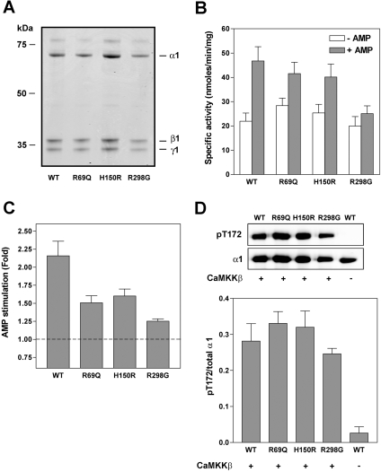
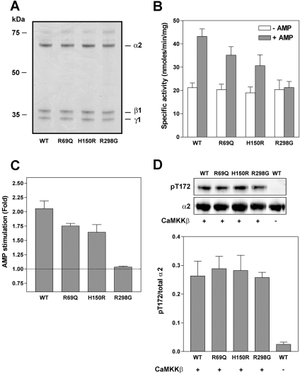
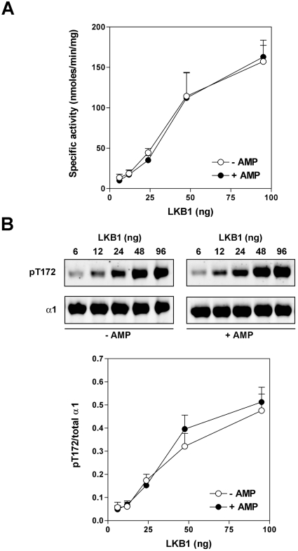
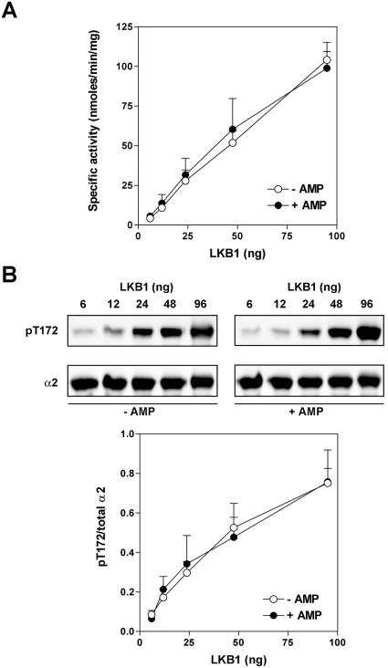
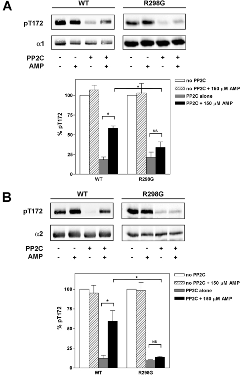
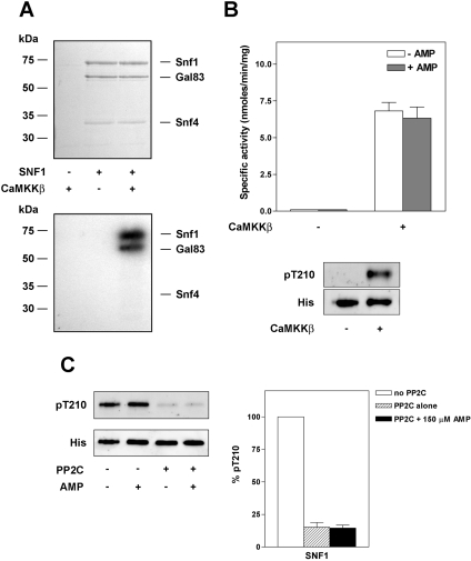
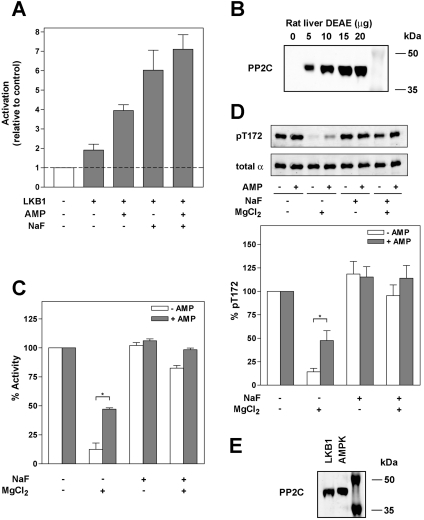
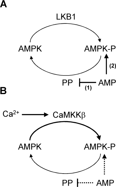
AMPK (AMP-activated protein kinase) is activated allosterically by AMP and by phosphorylation of Thr172 within the catalytic alpha subunit. Here we show that mutations in the regulatory gamma subunit reduce allosteric activation of the kinase by AMP. In addition to its allosteric effect, AMP significantly reduces the dephosphorylation of Thr172 by PP (protein phosphatase)2Calpha. Moreover, a mutation in the gamma subunit almost completely abolishes the inhibitory effect of AMP on dephosphorylation. We were unable to detect any effect of AMP on Thr172 phosphorylation by either LKB1 or CaMKKbeta (Ca2+/calmodulin-dependent protein kinase kinase beta) using recombinant preparations of the proteins. However, using partially purified AMPK from rat liver, there was an apparent AMP-stimulation of Thr172 phosphorylation by LKB1, but this was blocked by the addition of NaF, a PP inhibitor. Western blotting of partially purified rat liver AMPK and LKB1 revealed the presence of PP2Calpha in the preparations. We suggest that previous studies reporting that AMP promotes phosphorylation of Thr172 were misinterpreted. A plausible explanation for this effect of AMP is inhibition of dephosphorylation by PP2Calpha, present in the preparations of the kinases used in the earlier studies. Taken together, our results demonstrate that AMP activates AMPK via two mechanisms: by direct allosteric activation and by protecting Thr172 from dephosphorylation. On the basis of our new findings, we propose a simple model for the regulation of AMPK in mammalian cells by LKB1 and CaMKKbeta. This model accounts for activation of AMPK by two distinct signals: a Ca2+-dependent pathway, mediated by CaMKKbeta and an AMP-dependent pathway, mediated by LKB1.


Figures

Figure 1
Figure 1. Effect of mutations within γ subunit on allosteric activation of α1β1γ1 by AMP
Wild-type (WT) AMPK complex (α1β1γ1) or complexes harbouring point mutations in the γ subunit (R69Q, H150R or R298G) were expressed in E. coli and purified by nickel–Sepharose chromatography. (A) Proteins (∼1 μg) were resolved by SDS/PAGE and detected by staining with Coomassie Blue. (B) and (C) Following phosphorylation by CaMKKβ, AMPK activity of the complexes was determined using the SAMS peptide assay in the presence or absence of 150 μM AMP. Results are the means±S.E.M. for four independent experiments and are shown as the specific activity of the kinase (nmol of phosphate incorporated/min per mg of complex) in (B) and the fold stimulation in (C). (D) Western blot analysis of AMPK complexes probed with an antibody against either phospho-Thr172 (pT172) or α1. A representative blot is shown in the upper panel and the bottom panel shows the relative quantitation of pT172:total α1 as means±S.E.M for four independent experiments. Wild-type AMPK that had not been incubated with CaMKKβ is included as a negative control.
Figure 2
Figure 2. Effect of mutations within γ subunit on the allosteric activation of α2β1γ1 by AMP
The same set of experiments as described in the legend for Figure 1 were carried out using α2 complexes.
Figure 3
Figure 3. Effect of AMP on phosphorylation of α1β1γ1 by LKB1
(A) Wild-type α1β1γ1 was incubated with MgATP and varying amounts of LKB1 in the presence (closed circles) or absence (open circles) of 150 μM AMP for 10 min at 37 °C. At the end of the incubation an aliquot (5 μl) was removed, diluted in buffer and the AMPK activity was determined using the SAMS peptide assay. Note that in the SAMS peptide assay, AMP (150 μM final concentration) is present irrespective of whether AMP was included in the initial incubation with LKB1. (B) The top panel shows a representative Western blot of phospho-Thr172 (pT172) and total α1 of aliquots (5 μl) of the reactions described above. The bottom panel shows the relative quantitation of pT172:total α1 as means±S.E.M for three independent experiments.
Figure 4
Figure 4. Effect of AMP on phosphorylation of α2β1γ1 by LKB1
The same set of experiments as described in the legend for Figure 3 were carried out using α2 complexes.
Figure 5
Figure 5. Effect of AMP on dephosphorylation of recombinant AMPK by PP2C
Following phosphorylation of recombinant AMPK complexes (containing either wild-type γ1 or the R298G mutant) by CaMKKβ, aliquots (5 μl) were removed and incubated in the presence or absence of PP2C (26 ng) and presence or absence of AMP (150 μM) for 20 min at 37 °C. At the end of the incubation the reaction mixture was resolved by SDS/PAGE and analysed by Western blotting using anti-(phospho-Thr172) (pT172) and anti-α1 (A) or anti-(phospho-Thr172) and anti-α2 (B) antibodies. In each case a representative blot is shown in the top panels. In the bottom panels, Thr172 phosphorylation relative to a control (absence of PP2C and AMP) is shown as the means±S.E.M. for four independent experiments. Significant differences in Thr172 phosphorylation are denoted by *P<0.05). NS, not significant.
Figure 6
Figure 6. Effect of AMP on recombinant SNF1 complex
SNF1 (Snf1, Snf4, Gal83) was expressed in E. coli and purified on nickel–Sepharose, followed by gel-filtration chromatography. (A) Purified SNF1 was incubated with [32P]-ATP in the presence or absence of CaMKKβ and resolved by SDS/PAGE. The proteins were visualized by staining with Coomassie Blue (upper panel) and radiolabelled products detected by autoradiography (lower panel). (B) SNF1 activity was determined by the SAMS peptide assay in the presence or absence of 150 μM AMP. Activities are plotted as nmol of phosphate incorporated/min per mg of SNF1, and are the means±S.E.M for three independent experiments. Also shown is a representative blot of the reaction mixture probed with anti-(phospho-Thr172) (to detect phospho-Thr210 in Snf1) or anti-His6 antibody (to detect total Snf1). (C) SNF1 phosphorylated by CaMKKβ was incubated in the presence or absence of PP2C (26 ng) and the presence or absence of 150 μM AMP for 20 min at 37 °C. At the end of the incubation proteins were analysed by Western blotting using either anti-(phospho-Thr172) or anti-His6 antibody as described in the legend for Figure 5. The results are shown as means±S.E.M. for four independent experiments.
Figure 7
Figure 7. Inactivation of rat liver AMPK by endogenous PP activity
(A) Following dialysis into buffer lacking PP inhibitors, rat liver AMPK was incubated for 10 min at 37 °C in the presence of MgATP and the presence or absence of LKB1 (16 ng), AMP (150 μM) and/or NaF (50 mM). Following incubation, AMPK activity was determined. Results shown are plotted relative to AMPK activity in the presence of MgATP alone and are the means±S.E.M. for three independent experiments. (B) Partially purified rat liver AMPK extract was immunoblotted with an antibody against PP2Cα. (C) Rat liver AMPK was incubated in the presence or absence of MgCl2 (2.5 mM) and in the presence or absence of either 50 mM NaF, 150 μM AMP or both for 10 min at 37 °C. Following incubation, aliquots (5 μl) were removed and AMPK activity was determined using the SAMS peptide assay in the presence of 150 μM AMP (final concentration). (D) In parallel, aliquots (5 μl) were blotted with anti-(phospho-Thr172) or a mixture of anti-α1 and -α2 antibodies. A representative blot is shown (upper panel) and the lower panel shows the quantification of the relative level of Thr172 phosphorylation for three independent experiments (means±S.E.M.). *Significant differences (P<0.05) in AMPK activity or Thr172 phosphorylation. (E) Rat liver AMPK and LKB1, purified up to the Q-Sepharose step (as described in [29]), were probed with an anti-PP2Cα antibody.
Scheme 1
Scheme 1. Model for the regulation of AMPK
(A) AMP-dependent activation: under conditions that lead to an increase in AMP, dephosphorylation of AMPK is inhibited (mechanism 1). The identity of the PP responsible for dephosphorylation of AMPK in vivo is unknown. Since LKB1 is constitutively active, inhibition of the dephosphorylation reaction leads to an increase in Thr172 phosphorylation and activation of AMPK. In addition to increasing Thr172 phosphorylation, AMP allosterically activates AMPK (mechanism 2). (B) Ca2+-dependent activation: signals that increase Ca2+ activate CaMKKβ, increasing Thr172 phosphorylation and activation of AMPK, and this can occur without an increase in AMP. However, it is possible that in some situations both Ca2+ and AMP may increase in parallel, and under these conditions AMP will allosterically activate AMPK and inhibit dephosphorylation (denoted by the dashed lines).

References

    1. Carling D. The AMP-activated protein kinase cascade: a unifying system for energy control. Trends Biochem. Sci. 2004;29:18–24. - PubMed
    1. Kahn B. B., Alquier T., Carling D., Hardie D. G. AMP-activated protein kinase: ancient energy gauge provides clues to modern understanding of metabolism. Cell Metab. 2005;1:15–25. - PubMed
    1. Hardie D. G., Carling D., Carlson M. The AMP-activated/SNF1 protein kinase subfamily: metabolic sensors of the eukaryotic cell? Annu. Rev. Biochem. 1998;67:821–855. - PubMed
    1. Hong S. P., Leiper F. C., Woods A., Carling D., Carlson M. Activation of yeast Snf1 and mammalian AMP-activated protein kinase by upstream kinases. Proc. Natl. Acad. Sci. U.S.A. 2003;100:8839–8843. - PMC - PubMed
    1. Sutherland C. M., Hawley S. A., McCartney R. R., Leech A., Stark M. J. R., Schmidt M. C., Hardie D. G. Elm1p is one of three upstream kinases for the Saccharomyces cerevisiae SNF1 complex. Curr. Biol. 2003;13:1299–1305. - PubMed

Publication types