. 2007 Jan 22;274(1607):183-97.
doi: 10.1098/rspb.2006.3705.

Soft tissue and cellular preservation in vertebrate skeletal elements from the Cretaceous to the present

Affiliations

Soft tissue and cellular preservation in vertebrate skeletal elements from the Cretaceous to the present

Mary Higby Schweitzer et al. Proc Biol Sci. .

Erratum in

  • Proc Biol Sci. 2007 Dec 22;274(1629):3183

Abstract

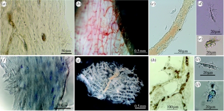
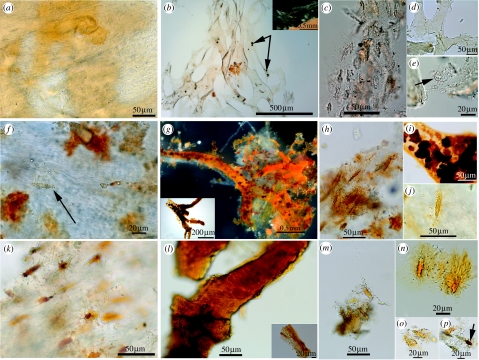
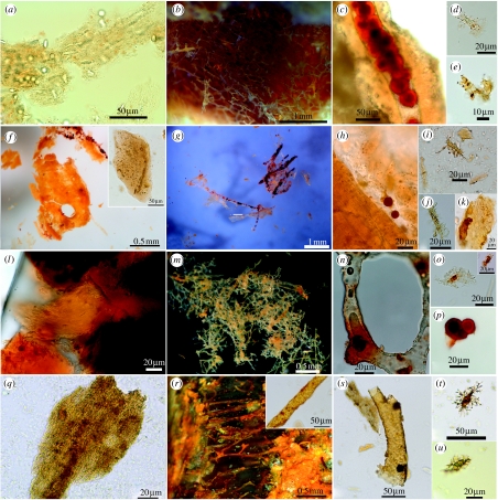
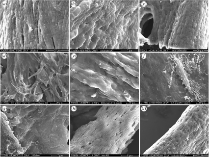
Soft tissues and cell-like microstructures derived from skeletal elements of a well-preserved Tyrannosaurus rex (MOR 1125) were represented by four components in fragments of demineralized cortical and/or medullary bone: flexible and fibrous bone matrix; transparent, hollow and pliable blood vessels; intravascular material, including in some cases, structures morphologically reminiscent of vertebrate red blood cells; and osteocytes with intracellular contents and flexible filipodia. The present study attempts to trace the occurrence of these four components in bone from specimens spanning multiple geological time periods and varied depositional environments. At least three of the four components persist in some skeletal elements of specimens dating to the Campanian. Fibrous bone matrix is more altered over time in morphology and less likely to persist than vessels and/or osteocytes. Vessels vary greatly in preservation, even within the same specimen, with some regions retaining pliability and other regions almost crystalline. Osteocytes also vary, with some retaining long filipodia and transparency, while others present with short and stubby filipodia and deeply pigmented nuclei, or are pigmented throughout with no nucleus visible. Alternative hypotheses are considered to explain the origin/source of observed materials. Finally, a two-part mechanism, involving first cross-linking of molecular components and subsequent mineralization, is proposed to explain the surprising presence of still-soft elements in fossil bone. These results suggest that present models of fossilization processes may be incomplete and that soft tissue elements may be more commonly preserved, even in older specimens, than previously thought. Additionally, in many cases, osteocytes with defined nuclei are preserved, and may represent an important source for informative molecular data.


Figures

Figure 1
Figure 1
Tissue and cells from extant ostrich and emu. (ae) Emu. (a) Fibrous texture of demineralized cortical bone matrix. Osteocytes and vessel channels are visible. (b) Vessels emerging from demineralized cortical bone fragment, after partial enzyme digestion. Pigmented blood products fill vessel lumen. (c) Isolated vessel shows intimate association with osteocytes. (d,e) Isolated osteocytes with radiating filipodia, liberated after collagenase digestion of cortical bone. Variation in degree of pigmentation is seen, as well as variation in overall cell shape. (fj) Ostrich. (f) Histochemical staining emphasizes fibrous texture of demineralized ostrich cortical bone matrix. Osteocytes are interspersed with collagen fibres. (g) Interconnecting lattice of vessels liberated from fresh ostrich cortical bone after demineralization and enzymatic digestion as above. (h) Ostrich vessel and associated matrix with osteocytes after demineralization and digestion. Blood breakdown products fill vessel lumen. (i) Unstained, isolated osteocyte is elongate with extensive filipodia. (j) Cell body is expanded and has been stained to reveal cellular detail. All specimens derived from 14+ year post-mortem specimen except (g), from ca 1 year post-mortem. Scale bars are as indicated.
Figure 2
Figure 2
Tissue and cells: moa, mammoth, and mastodon. (MOR OFT255, ae) Moa. (a) Fibrous texture, embedded osteocytes and natural pigmentation of demineralized cortical bone. Vessels are not apparent in this image. (b) Interconnecting network of isolated, transparent vessels, with dark-bodied fungal spores visible within vessel lumen (arrows). Inset shows vessels emerging from digesting bone fragment. (c) Enzyme-resistant tissue associated with osteocyte cell bodies. (d) Isolated fragment of vessel shows natural pigmentation. (e) Transparent, round-bodied osteocyte (arrow) with radiating filipodia, still closely associated or attached to remnants of fibrous matrix. (MOR 604, fj) Mammoth. (f) Demineralized mammoth skull fragment shows uneven pigmentation and osteocyte (arrow) with filipodia within fibrous matrix. (g) Flexible, deeply pigmented vessels with associated matrix (white) sheet material. Vascular contents appear as deeply pigmented microstructures. Inset shows vessel with similar pigmentation and internal microstructures, but crystalline, not flexible. (h) Flexible, fibrous matrix containing multiple pigmented osteocytes with interconnecting filipodia. (i) Vessel shows consistent wall thickness and intravascular material. (j) Orange-pigmented osteocyte with filipodia radiating outward into fibrous matrix. (MOR 605, kp) Mastodon. (k) Fibrous texture of demineralized matrix encloses abundant, naturally pigmented osteocytes, with distinct nuclei and long filipodia extending into matrix fibres. (l) Fragmented and deeply pigmented crystalline vessel. Inset shows another vessel fragment with crystalline surface structure. (mp) Osteocytes with interconnected filipodia, some showing distinct nuclei (p, arrow). Scale bars are as indicated.
Figure 3
Figure 3
Soft tissue and cells from selected dinosaur specimens. (MOR 555, ae) Tyrannosaurus rex. (a) Fragment of demineralized, flexible cortical bone matrix with fibrous texture. Transparent osteocytes are visible among the matrix fibres. (b) Interconnected network of transparent and/or lightly pigmented vessels, released after demineralization. Vessels are hollow and supple. (c) Vessel fragment filled with small red microstructures with opaque centres, surrounded by fibrous bone matrix containing osteocytes. Pigmentation is natural. (d) Internal, intracellular contents are visible in isolated osteocyte with filipodia. (e) Filipodia associated with small fragment of fibrous matrix. (MOR 699, fk) Triceratops horridus. (f) Deeply pigmented, flexible matrix tissue with vascular channel in centre. Inset shows fibrous matrix with pigmented spaces representing retained osteocytes and partial vessel. (g) Isolated transparent vessels containing darkly pigmented intravascular structures. Vessels were rare in this specimen. (h) Round red microstructures associated with longitudinal space, possibly representing vascular canal running through demineralized fibrous matrix. (i,j) Isolated osteocytes with long filipodia. (k) Vessel fragment showing surface texture and natural pigmentation. (MOR 1125, lp) Tyrannosaurus rex. (l) Fragment of demineralized, naturally pigmented fibrous matrix tissue containing osteocytes. (m) Interconnected, flexible, transparent vessels with pigmented intravascular structures with varying diameters similar to extant vessels. (n) Vessel with hollow lumen and uniformly thick vessel wall, containing deeply pigmented intravascular microstructure. (o) Isolated, free-floating osteocytes, with long, extensive filipodia and intracellular contents. (p) Three naturally pigmented intravascular microstructures with opaque centre, expressed from vessel. (MOR 794, qu) Brachylophosaur canadensis. (q) Flexible, fibrous demineralized bone matrix with variable pigment distribution. (r) Orange-pigmented vessels, either crystalline or flexible. Flexible vessel walls, less transparent than in other dinosaurs (Inset). (s) Isolated, flexible branching vessel with intravascular contents. (t,u) Naturally pigmented osteocytes with extensive three-dimensional filipodial network. Scale bars are as indicated.
Figure 4
Figure 4
SEM of demineralized bone matrix from multiple specimens. (a) Fresh chicken showing fibre bundles. Crossbanding is not within SEM resolution capability. (b) Emu matrix, with fibre bundles at higher magnification. (c) Moa trabecular bone (MOR OFT255). (d) Mammoth (MOR 91.72). (e) Mammoth (MOR 604). (f) Mastodon (MOR 605). Featureless matrix is impossible to image without also imaging osteocytes (centre). (g) Tyrannosaurus rex (MOR 555). (h) Tyrannosaurus rex (FMNH-PR-2081). (i) Theropod indeterminant (MN 4802-V). Magnifications and scale bars are as indicated.

References

    1. Adams J.S, Organ C. Histologic determination of ontogenetic patterns and processes in hadrosaurian ossified tendons. J. Vert. Paleontol. 2005;25:614–622. doi: 10.1671/0272-4634(2005)025[0614:HDOOPA]2.0.CO;2 - DOI
    1. Allison P.A. The role of anoxia in the decay and mineralization of proteinaceous macro-fossils. Paleobiology. 1988;14:139–154.
    1. Ambler R.P, Daniel M. Proteins and molecular paleontology. Phil. Trans. R. Soc. B. 1991;333:381–389. - PubMed
    1. Bada J.L, Wang X.Y.S, Hamilton H. Preservation of key biomolecules in the fossil record: current knowledge and future challenges. Phil. Trans. R. Soc. B. 1999;354:77–86. doi:10.1098/rstb.1999.0361 - DOI - PMC - PubMed
    1. Behrensmeyer A.K, Kidwell S.M. Taphonomy's contributions to paleobiology. Paleobiology. 1985;11:105–119.

Publication types

LinkOut - more resources