

Hypervariability within the Rifin, Stevor and Pfmc-2TM superfamilies in Plasmodium falciparum
- PMID: 17148488
- PMCID: PMC1751529
- DOI: 10.1093/nar/gkl942
Hypervariability within the Rifin, Stevor and Pfmc-2TM superfamilies in Plasmodium falciparum
Abstract
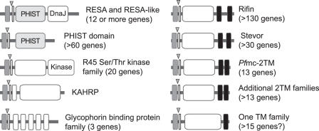
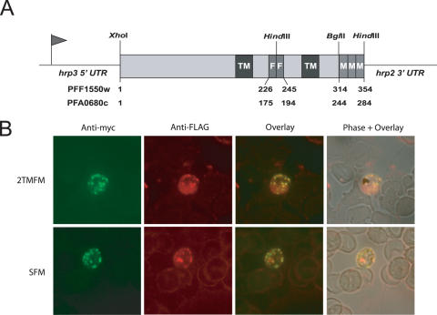
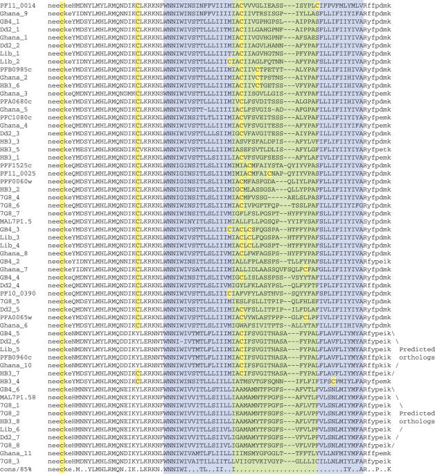
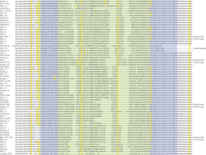
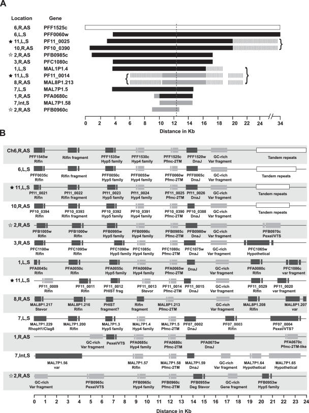
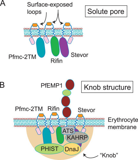
The human malaria parasite, Plasmodium falciparum, possesses a broad repertoire of proteins that are proposed to be trafficked to the erythrocyte cytoplasm or surface, based upon the presence within these proteins of a Pexel/VTS erythrocyte-trafficking motif. This catalog includes large families of predicted 2 transmembrane (2TM) proteins, including the Rifin, Stevor and Pfmc-2TM superfamilies, of which each possesses a region of extensive sequence diversity across paralogs and between isolates that is confined to a proposed surface-exposed loop on the infected erythrocyte. Here we express epitope-tagged versions of the 2TM proteins in transgenic NF54 parasites and present evidence that the Stevor and Pfmc-2TM families are exported to the erythrocyte membrane, thus supporting the hypothesis that host immune pressure drives antigenic diversity within the loop. An examination of multiple P.falciparum isolates demonstrates that the hypervariable loop within Stevor and Pfmc-2TM proteins possesses sequence diversity across isolate boundaries. The Pfmc-2TM genes are encoded within large amplified loci that share profound nucleotide identity, which in turn highlight the divergences observed within the hypervariable loop. The majority of Pexel/VTS proteins are organized together within sub-telomeric genome neighborhoods, and a mechanism must therefore exist to differentially generate sequence diversity within select genes, as well as within highly defined regions within these genes.
Figures
Similar articles
-
Temporal expression and localization patterns of variant surface antigens in clinical Plasmodium falciparum isolates during erythrocyte schizogony.PLoS One. 2012;7(11):e49540. doi: 10.1371/journal.pone.0049540. Epub 2012 Nov 15. PLoS One. 2012. PMID: 23166704 Free PMC article.
-
A comparative study of the localization and membrane topology of members of the RIFIN, STEVOR and PfMC-2TM protein families in Plasmodium falciparum-infected erythrocytes.Malar J. 2015 Jul 16;14:274. doi: 10.1186/s12936-015-0784-2. Malar J. 2015. PMID: 26173856 Free PMC article.
-
Expression switching in the stevor and Pfmc-2TM superfamilies in Plasmodium falciparum.Mol Microbiol. 2007 Jun;64(6):1621-34. doi: 10.1111/j.1365-2958.2007.05767.x. Mol Microbiol. 2007. PMID: 17555442
-
The varieties of gene amplification, diversification and hypervariability in the human malaria parasite, Plasmodium falciparum.Mol Biochem Parasitol. 2009 Aug;166(2):109-16. doi: 10.1016/j.molbiopara.2009.04.003. Epub 2009 Apr 16. Mol Biochem Parasitol. 2009. PMID: 19375460 Review.
-
Variable Surface Antigens of Plasmodium falciparum: Protein Families with Divergent Roles.Protein Pept Lett. 2024;31(6):409-423. doi: 10.2174/0109298665298567240530170924. Protein Pept Lett. 2024. PMID: 38910420 Review.
Cited by
-
Temporal expression and localization patterns of variant surface antigens in clinical Plasmodium falciparum isolates during erythrocyte schizogony.PLoS One. 2012;7(11):e49540. doi: 10.1371/journal.pone.0049540. Epub 2012 Nov 15. PLoS One. 2012. PMID: 23166704 Free PMC article.
-
Plasmodium falciparum STEVOR proteins are highly expressed in patient isolates and located in the surface membranes of infected red blood cells and the apical tips of merozoites.Infect Immun. 2008 Jul;76(7):3329-36. doi: 10.1128/IAI.01460-07. Epub 2008 May 12. Infect Immun. 2008. PMID: 18474651 Free PMC article.
-
Molecular Players at the Sorting Stations of Malaria Parasite 'Plasmodium falciparum'.Curr Protein Pept Sci. 2024;25(6):427-437. doi: 10.2174/0113892037282522240130090156. Curr Protein Pept Sci. 2024. PMID: 38409726 Review.
-
Absence of erythrocyte sequestration and lack of multicopy gene family expression in Plasmodium falciparum from a splenectomized malaria patient.PLoS One. 2009 Oct 14;4(10):e7459. doi: 10.1371/journal.pone.0007459. PLoS One. 2009. PMID: 19826486 Free PMC article.
-
'2TM proteins': an antigenically diverse superfamily with variable functions and export pathways.PeerJ. 2018 May 11;6:e4757. doi: 10.7717/peerj.4757. eCollection 2018. PeerJ. 2018. PMID: 29770278 Free PMC article.
References
-
- Haeggstrom M., Kironde F., Berzins K., Chen Q., Wahlgren M., Fernandez V. Common trafficking pathway for variant antigens destined for the surface of the Plasmodium falciparum-infected erythrocyte. Mol. Biochem. Parasitol. 2004;133:1–14. - PubMed
-
- Papakrivos J., Newbold C.I., Lingelbach K. A potential novel mechanism for the insertion of a membrane protein revealed by a biochemical analysis of the Plasmodium falciparum cytoadherence molecule PfEMP-1. Mol. Microbiol. 2005;55:1272–1284. - PubMed
-
- Taraschi T.F., O'Donnell M., Martinez S., Schneider T., Trelka D., Fowler V.M., Tilley L., Moriyama Y. Generation of an erythrocyte vesicle transport system by Plasmodium falciparum malaria parasites. Blood. 2003;102:3420–3426. - PubMed
-
- Trelka D.P., Schneider T.G., Reeder J.C., Taraschi T.F. Evidence for vesicle-mediated trafficking of parasite proteins to the host cell cytosol and erythrocyte surface membrane in Plasmodium falciparum infected erythrocytes. Mol. Biochem. Parasitol. 2000;106:131–145. - PubMed
Publication types
MeSH terms
Substances
Grants and funding
LinkOut - more resources
Full Text Sources
Other Literature Sources
