Review
. 2006 Dec;169(6):1901-9.
doi: 10.2353/ajpath.2006.060681.

Molecular basis of epithelial barrier regulation: from basic mechanisms to clinical application

Affiliations
Review

Molecular basis of epithelial barrier regulation: from basic mechanisms to clinical application

Jerrold R Turner. Am J Pathol. 2006 Dec.

Abstract

The intestinal epithelium is faced with the complex task of providing a barrier while also allowing nutrient and water absorption. The frequency with which these processes are disrupted in disease can be taken as evidence of their importance. It is therefore of interest to define the mechanisms of altered intestinal barrier and transport function and develop means to correct disease-associated defects. Over the past 10 years, some of the molecular events underlying physiological epithelial barrier regulation have been described. Remarkably, recent advances have shown that activation of the same mechanisms is central to barrier dysfunction in both in vitro and in vivo models of disease. Although the contribution of barrier dysfunction to pathogenesis of chronic disease remains incompletely understood, it is now clear that cytoskeletal regulation of barrier function is both an important pathogenic process and that targeted inhibition of myosin light chain kinase, which affects this cytoskeleton-dependent tight junction dysfunction, is an attractive candidate for therapeutic intervention.


Figures
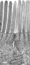
Figure 1
Figure 1
The epithelial tight junction. Electron micrograph of the junction between two adjacent villus absorptive enterocytes. Note the actin core of the microvilli that extends into a filamentous mesh, the cortical actin web, within the apical cytoplasm. Denser filament accumulations are apparent surrounding the apical junction complex. The latter is composed of the tight junction, a zone of close apposition of adjacent plasma membranes just beneath the apical surface, and the adherens junction, which is immediately sub apical to the tight junction. Bar = 200 nm.
Figure 2
Figure 2
Actin depolymerization induces caveolae-mediated occludin endocytosis. Three-dimensional projections of monolayers labeled for occludin (red) and caveolin-1 (green), a marker of caveolae, are shown. Rather than the ordered appearance of occludin encircling the apical portion of the cell with few intracellular occludin-continuing vesicles in control epithelia (A), abundant intracellular occludin-continuing vesicles are evident after actin depolymerization (B). Many of these newly formed vesicles also contain caveolin-1, as apparent from the yellow colocalization signal. Bar = 5 μm.
Figure 3
Figure 3
In vivo T-cell activation causes occludin endocytosis. Three hours following systemic T-cell activation, mouse jejunum was snap frozen and fluorescently labeled for occludin (red), F-actin (green), and nuclei (blue). In addition to some residual tight junction-localized occludin, which is associated with the perijunctional actomyosin ring, bright vesicular occludin deposits are present within the cytoplasm of these villus enterocytes. Intracellular occludin deposits were not seen within the cytoplasm of villus enterocytes from control animals. Bar = 5 μm.
Figure 4
Figure 4
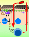
A mechanism-based model of intestinal disease. This model proposes a self-amplifying pathway where a small amount of luminal, eg, bacterial-derived, molecules cross the epithelium to activate lamina propria immune cells. This results in secretion of proinflammatory cytokines, eg, TNF, that signal to intestinal epithelial. This TNF receptor 2 (TNFR2)-mediated signal increases both MLCK transcription and enzymatic activity, resulting in occludin endocytosis and epithelial barrier dysfunction. The barrier loss results in greater access leakage of luminal material, greater immune activation, and even greater barrier defects. In the absence of appropriate regulatory cues, this triggers a self-amplifying cascade. Although described here as being initiated by luminal molecules activating immune cells, it should be apparent that the primary event could as easily be inappropriate immune activation or epithelial barrier dysfunction.

Similar articles

Cited by

References

    1. Sandler RS, Everhart JE, Donowitz M, Adams E, Cronin K, Goodman C, Gemmen E, Shah S, Avdic A, Rubin R. The burden of selected digestive diseases in the United States. Gastroenterology. 2002;122:1500–1511. - PubMed
    1. Clayburgh DR, Shen L, Turner JR. A porous defense: the leaky epithelial barrier in intestinal disease. Lab Invest. 2004;84:282–291. - PubMed
    1. Hill GR, Ferrara JL. The primacy of the gastrointestinal tract as a target organ of acute graft-versus-host disease: rationale for the use of cytokine shields in allogeneic bone marrow transplantation. Blood. 2000;95:2754–2759. - PubMed
    1. Baert FJ, D’Haens GR, Peeters M, Hiele MI, Schaible TF, Shealy D, Geboes K, Rutgeerts PJ. Tumor necrosis factor alpha antibody (infliximab) therapy profoundly down-regulates the inflammation in Crohn’s ileocolitis. Gastroenterology. 1999;116:22–28. - PubMed
    1. Suenaert P, Bulteel V, Lemmens L, Noman M, Geypens B, Van Assche G, Geboes K, Ceuppens JL, Rutgeerts P. Anti-tumor necrosis factor treatment restores the gut barrier in Crohn’s disease. Am J Gastroenterol. 2002;97:2000–2004. - PubMed

Publication types

MeSH terms