. 2006 Dec;169(6):2042-53.
doi: 10.2353/ajpath.2006.060081.

Resistin as an intrahepatic cytokine: overexpression during chronic injury and induction of proinflammatory actions in hepatic stellate cells

Affiliations

Resistin as an intrahepatic cytokine: overexpression during chronic injury and induction of proinflammatory actions in hepatic stellate cells

Cristiana Bertolani et al. Am J Pathol. 2006 Dec.

Abstract

Obesity and insulin resistance accelerate the progression of fibrosis during chronic liver disease. Resistin antagonizes insulin action in rodents, but its role in humans is still controversial. The aims of this study were to investigate resistin expression in human liver and to evaluate whether resistin may affect the biology of activated human hepatic stellate cells (HSCs), key modulators of hepatic fibrogenesis. Resistin gene expression was low in normal human liver but was increased in conditions of severe fibrosis. Up-regulation of resistin during chronic liver damage was confirmed by immunohistochemistry. In a group of patients with alcoholic hepatitis, resistin expression correlated with inflammation and fibrosis, suggesting a possible action on HSCs. Exposure of cultured HSCs to recombinant resistin resulted in increased expression of the proinflammatory chemokines monocyte chemoattractant protein-1 and interleukin-8, through activation of nuclear factor (NF)-kappaB. Resistin induced a rapid increase in intracellular calcium concentration, mainly through calcium release from intracellular inositol triphosphate-sensitive pools. The intracellular calcium chelator BAPTA-AM blocked resistin-induced NF-kappaB activation and monocyte chemoattractant protein-1 expression. In conclusion, this study shows a role for resistin as an intrahepatic cytokine exerting proinflammatory actions in HSCs, via a Ca2+/NF-kappaB-dependent pathway and suggests involvement of this adipokine in the pathophysiology of liver fibrosis.


Figures

Figure 1
Figure 1
Resistin gene expression in human liver tissue. Expression of resistin mRNA was analyzed by real-time PCR, as described in Materials and Methods, in normal human liver tissues (NHL, n = 4) and in liver tissue of patients with end-stage fibrosis (LF, n = 5). mRNA isolated from bone marrow (BM) was used as a positive control for resistin expression. Resistin mRNA (mean + SD) is expressed as 2−ΔCt (ΔCt = Ct of the target gene minus Ct of the housekeeping gene). *P < 0.05 versus NHL.
Figure 2
Figure 2
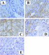
Immunohistochemistry for resistin in human liver tissue. Representative photomicrographs of resistin immunostaining (brown color) in specimens from patients with normal liver (A), chronic hepatitis C (B), alcohol-induced liver disease (C), and nonalcoholic steatohepatitis (D). E: A negative control slide (alcohol-induced liver disease). Original magnifications, ×400.
Figure 3
Figure 3
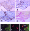
Comparison of resistin expression with that of specific cellular markers or collagen deposition. A–D: Serial sections of liver tissue from a patient with severe alcohol-induced liver disease were analyzed by immunohistochemistry for resistin (A), CD43 (inflammatory cells, B), α-SMA (fibrogenic myofibroblasts, C), or stained with Sirius Red (collagen deposition, D). E–G: Liver tissue from a patient with severe alcohol-induced liver disease was analyzed by immunofluorescence for resistin (E, green) or CD43 (F, red). G: Merged images, with double-positive cells indicated by arrows. Original magnifications: ×200 (A–D); ×800 (E–G).
Figure 4
Figure 4
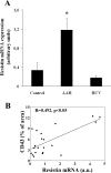
Up-regulated resistin expression in patients with AAH. A: Intrahepatic gene expression of resistin (mean + SD), as assessed by quantitative PCR, in patients with AAH (n = 23) or with chronic HCV-related hepatitis (HCV, n = 14), and controls (n = 6). 18S rRNA was used as the housekeeping gene. Resistin mRNA values are given as arbitrary units. *P < 0.001 versus control livers. B: Correlation between intrahepatic resistin mRNA expression and CD43-positive area in the group of patients with AAH (n = 23). In two patients, data for correlation were not available.
Figure 5
Figure 5
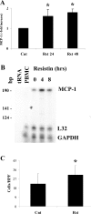
Resistin stimulates MCP-1 expression in HSCs. A: Serum-starved HSCs were exposed for 24 or 48 hours to 500 ng/ml resistin, as indicated. At the end of incubation, conditioned medium was collected, and MCP-1 concentrations were measured by ELISA. *P ≤ 0.05 versus control. Data are expressed as mean ± SE of three independent experiments. B: Serum-starved HSCs were stimulated with 250 ng/ml resistin for the indicated time points. Total RNA was analyzed for MCP-1 gene expression by RNase protection assay, as described in Materials and Methods. Transfer RNA (tRNA) and peripheral blood mononuclear cell RNA (PBMC) were used as negative and positive controls for hybridization, respectively. C: HSCs were incubated in the presence or absence of 500 ng/ml resistin for 48 hours. Aliquots of cell supernatant were analyzed in chemotactic assays using human peripheral blood mononuclear cells, as described in Materials and Methods. The number of cells migrating to the underside of the filters was quantified in 10 high-power fields (HPFs). Results are the mean ± SE of four different experiments.
Figure 6
Figure 6
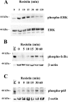
Resistin activates different intracellular signaling pathways in HSCs. A: Serum-deprived HSCs were exposed to 500 ng/ml resistin for different time points, as indicated. Ten μg of total cell lysate were separated by SDS-PAGE and sequentially immunoblotted with antibodies specifically recognizing the phosphorylated form of ERK (top) or total ERK (bottom). B: Serum-deprived HSCs were incubated with 500 ng/ml resistin at the indicated time points. Total cell lysates were separated by SDS-PAGE and analyzed by immunoblotting using antibodies specifically recognizing phosphorylated IκBα (top) or β-actin (bottom). A lysate from HepG2 cells exposed to 200 ng/ml TNF-α for 15 minutes was used as positive control (PC). C: The experiment was conducted exactly as described in B, but the membranes were blotted with antibodies against phosphorylated p65NF-κB (top) or β-actin (bottom). Migration of the molecular weight markers is shown on the left.
Figure 7
Figure 7
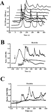
Effect of resistin on intracellular Ca2+ concentration. A: Subconfluent serum-starved HSCs were loaded with the fluorescent dye Fura-2AM. Intracellular Ca2+ concentration was measured by image analysis, as described in Materials and Methods. Resistin (100 ng/ml) was added at time 0. Each trace is the response measured in an individual cell at 3-second time intervals. B: The effects of resistin (100 ng/ml, added at time 0) were analyzed in control conditions (black squares), after depletion of intracellular calcium stores with 30 nmol/L thapsigargin (added at the arrow, black trace), or in the absence of extracellular calcium (open squares). Traces are the mean of at least seven cells. C: The effects of resistin (100 ng/ml, added at time 0) were analyzed in control conditions (black squares), after a 10-minute preincubation with 4 μmol/L U73122 (open diamonds) or after preincubation with 50 μmol/L 2-APB (black trace). Traces are the mean of at least seven cells.
Figure 8
Figure 8
NF-κB activation by resistin requires intracellular calcium. A: Serum-deprived HSCs were preincubated with 10 μmol/L BAPTA-AM or vehicle for 30 minutes, before the addition of 250 ng/ml resistin for 15 minutes, as indicated. Total cell lysates were separated by SDS-PAGE and analyzed by immunoblotting using antibodies specifically recognizing the phosphorylated form of IκBα. Total protein lysate from HepG2 cells exposed to 200 ng/ml TNF-α for 15 minutes was used as positive control. The membrane was stripped and reblotted with β-actin antibody to ensure equal loading. Migration of the molecular weight marker is shown on the left. B: Serum-starved HSCs were preincubated with BAPTA-AM for 10 minutes and then exposed to 250 ng/ml resistin. NF-κB-dependent gene expression was assessed by luciferase reporter gene assay as described in Materials and Methods. Results are shown as fold increase with respect to untreated cells. *P < 0.05 versus resistin alone.
Figure 9
Figure 9
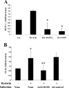
Resistin-induced chemokine secretion is NF-κB- and calcium-dependent. A: Serum-starved HSCs were preincubated either with the NF-κB inhibitory peptide SN50 (18 μmol/L) for 15 minutes or with BAPTA-AM (10 μmol/L) for 30 minutes, before exposure for 24 hours to 500 ng/ml resistin. At the end of incubation, MCP-1 concentration in the conditioned medium was measured by ELISA. Results are shown as fold increase with respect to untreated cells. *P < 0.05 versus resistin alone. B: HSCs were infected with an IκB-superrepressor adenovirus (Ad-IκB-SR) or control adenovirus (Ad-control), as described in Materials and Methods. After infection, serum-deprived HSCs were incubated with 250 ng/ml resistin for 24 hours, as indicated, and IL-8 concentration in the conditioned medium was measured by ELISA. Results are shown as fold increase with respect to untreated cells. *P < 0.05 versus buffer; **P < 0.05 versus Ad control + resistin.

References

    1. Fantuzzi G. Adipose tissue, adipokines, and inflammation. J Allergy Clin Immunol. 2005;115:911–919. - PubMed
    1. Day CP. From fat to inflammation. Gastroenterology. 2006;130:207–210. - PubMed
    1. Steppan CM, Brown EJ, Wright CM, Bhat S, Banerjee RR, Dai CY, Enders GH, Silberg DG, Wen X, Wu GD, Lazar MA. A family of tissue-specific resistin-like molecules. Proc Natl Acad Sci USA. 2001;98:502–506. - PMC - PubMed
    1. Steppan CM, Bailey ST, Bhat S, Brown EJ, Banerjee RR, Wright CM, Patel HR, Ahima RS, Lazar MA. The hormone resistin links obesity to diabetes. Nature. 2001;409:307–312. - PubMed
    1. Muse ED, Obici S, Bhanot S, Monia BP, McKay RA, Rajala MW, Scherer PE, Rossetti L. Role of resistin in diet-induced hepatic insulin resistance. J Clin Invest. 2004;114:232–239. - PMC - PubMed

Publication types