. 2006 Dec;169(6):2066-74.
doi: 10.2353/ajpath.2006.060211.

Oval cell response in 2-acetylaminofluorene/partial hepatectomy rat is attenuated by short interfering RNA targeted to stromal cell-derived factor-1

Affiliations

Oval cell response in 2-acetylaminofluorene/partial hepatectomy rat is attenuated by short interfering RNA targeted to stromal cell-derived factor-1

Donghang Zheng et al. Am J Pathol. 2006 Dec.

Abstract

Stromal cell-derived factor-1 (SDF-1) is known to play an essential role in the regulation of stem/progenitor cell trafficking. During hepatic stem, or oval, cell activation, SDF-1 has been reported to be up-regulated within the liver, implying a possible role in oval cell-aided liver regeneration. In the present study, SDF-1 expression was knocked down in the liver of 2-acetylaminofluorene/partial hepatectomy-treated rats using short interfering RNA delivered by recombinant adenovirus. The oval cell response was compromised in these animals, as evidenced by a decreased number of OV6-positive oval cells. In addition, knockdown of SDF-1 expression caused a dramatic decrease in alpha-fetoprotein expression, implying impaired oval cell activation in these animals. Terminal deoxynucleotidyl transferase-mediated dUTP nick-end-labeling assay showed no significant apoptosis related to SDF-1 suppression. Instead, as revealed by Ki67 immunohistochemistry, the suppression of SDF-1 resulted in decrease of hepatic cell proliferation, implying the repair process had been inhibited in these animals. These results indicate that SDF-1 is an essential molecule needed in oval cell activation.


Figures

Figure 1
Figure 1
Immunofluorescent staining of SDF-1 in normal rat liver and 2AAF/PHx rat liver. Normal liver (A) and 2AAF/PHx (B) rat, day 9. C: Primary antibody replaced by normal goat IgG (negative control) 2AAF/PHx rat, day 9. Arrows point to SDF-1-positive hepatocytes. SDF-1 (green) expression can be seen in most of the hepatocytes within the pericentral region on the liver of 2AAF/PHx rat. C: Notice that in this model, most cells within portal triads are SDF-1-negative. A: SDF-1 expression is not detected in normal liver. Autofluorescence is displayed as orange color under the dual-path fluorescein isothiocyanate-Texas Red filter used here. PT, portal triad; CV, central vein. Original magnification, ×20.
Figure 2
Figure 2
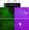
Ad-siSDF knocks down SDF-1 expression in 2AAF/PHx rat livers and inhibits the oval cell reaction in these animals. A: PCR detection of viral DNA in the livers day 9 after 2AAF/PHx and virus infusion. DNA from day-9 2AAF/PHx-treated liver was used as negative control. B and C: Liver sections from 2AAF/PHx rat treated with Ad-scramble, day 9. D and E: Liver sections from 2AAF/PHx rat treated with Ad-siSDF, day 9. B and D: Immunofluorescence of SDF-1 (green) counterstained with DAPI (blue) for nuclear. SDF-1 was knocked down in Ad-siSDF-treated animal liver (compare B with D). C and E: H&E staining of liver sections showing suppressed oval cell response in Ad-siSDF-treated animals (E) compared with control animal (C). Arrows in B and D point to SDF-1-positive hepatocytes. Arrows in C and E point to oval cells. Original magnification, ×20.
Figure 3
Figure 3
Knockdown of SDF-1 and decreased AFP expression after siSDF treatment. SDF-1 and AFP protein level in rat liver was detected by Western blot. Protein sample (10 μg) from the liver of each animal was loaded into each lane. Relative quantity of each band is determined by normalizing its total density to that of loading control in the same lane. Ad-siSDF-induced RNA interference results in considerable decrease in SDF-1 expression, accompanied by a decrease of AFP expression from these rat livers (P < 0.05, Student’s t-test).
Figure 4
Figure 4
OV6 immunostaining of rat liver sections. A: 2AAF/PHx rat treated with Ad-scramble, day 9. B: High magnification of the bracketed area in A. C: 2AAF/PHx rat treated with Ad-siSDF, day 9. D: High magnification of bracketed area in C. Less OV6-positive (brown) oval cells are seen in Ad-siSDF-treated animals (C and D) than in control animals (A and B). E: Comparison of the number of OV6-positive cells in different group of animals. OV6-positive oval cells were counted from 25 randomly selected fields (×40) in sections from each group. The number of oval cells per field was 88.9 ± 29.8 in control rat and 24.2 ± 17.4 in Ad-siSDF-treated rat (mean ± 2 SD, P < 0.01, Student’s t-test). Arrows point to oval cells. Original magnification: ×10 (A and C); ×40 (B and D).
Figure 5
Figure 5
No significant apoptosis detected by TUNEL staining in 2AAF/PHx rat livers. A: 2AAF/PHx rat, day 9. B: 2AAF/PHx/Ad-scramble-treated rat, day 9. C: 2AAF/PHx/Ad-siSDF-treated rat, day 9. Arrows point to apoptotic cells (green), which are sporadic on the liver sections from all animals. The extent of apoptosis in the liver is similar between all three groups at this time point. Original magnification, ×10.
Figure 6
Figure 6
Knockdown of SDF-1 hinders hepatic cell proliferation. A: 2AAF/PHx/Ad-scramble-treated rat, day 9. B: 2AAF/PHx/Ad-siSDF-treated rat, day 9. C: 2AAF/PHx/Ad-scramble-treated rat, day 13. D: 2AAF/PHx/Ad-siSDF-treated rat, day 13. Arrows point to proliferating oval cells and arrowheads point to proliferating hepatocytes. Original magnification, ×20. E: Comparison of the number of proliferating cells in different group of animals. Ki67-positive cells (brown nucleus) were counted as proliferating cells from 25 randomly selected fields in sections of each group. The cells residing within sinusoids were intentionally ignored because they were not related to oval cells. The number of proliferating cells per field (under ×20 magnification) was 388.7 ± 49.1 in control rat versus 106.4 ± 40.6 in Ad-siSDF-treated rat at day 9 and 465.8 ± 53.7 in control rat versus 157.5 ± 20.2 in Ad-siSDF-treated rat at day 13 (mean ± 2 SD, P < 0.01, Student’s t-test).

References

    1. Evarts RP, Nagy P, Marsden E, Thorgeirsson SS. A precursor-product relationship exists between oval cells and hepatocytes in rat liver. Carcinogenesis. 1987;8:1737–1740. - PubMed
    1. Tee LB, Kirilak Y, Huang WH, Morgan RH, Yeoh GC. Differentiation of oval cells into duct-like cells in preneoplastic liver of rat placed on a choline-deficient diet supplement with ethionine. Carcinogenesis. 1994;15:4116–4124. - PubMed
    1. Paku S, Schnur J, Nagy P, Thorgeirsson SS. Origin and structural evolution of the early proliferating oval cells in rat liver. Am J Pathol. 2001;158:1313–1323. - PMC - PubMed
    1. Petersen BE, Bowen WC, Patrene KD, Mars WM, Sullivan AK, Murase N, Boggs SS, Greenberger JS, Goff JP. Bone marrow as a potential source of hepatic oval cells. Science. 1999;284:1168–1170. - PubMed
    1. Ratajczak MZ, Kucia M, Reca R, Majka M, Janowska-Wieczorek A, Ratajczak J. Stem cell plasticity revisited: cXCR4-positive cells expressing mRNA for early muscle, liver and neural cells ‘hide out’ in the bone marrow. Leukemia. 2004;18:29–40. - PubMed

Publication types

LinkOut - more resources