. 2006 Dec;169(6):2161-70.
doi: 10.2353/ajpath.2006.060329.

Genetic ablation of vitamin D activation pathway reverses biochemical and skeletal anomalies in Fgf-23-null animals

Affiliations

Genetic ablation of vitamin D activation pathway reverses biochemical and skeletal anomalies in Fgf-23-null animals

Despina Sitara et al. Am J Pathol. 2006 Dec.

Abstract

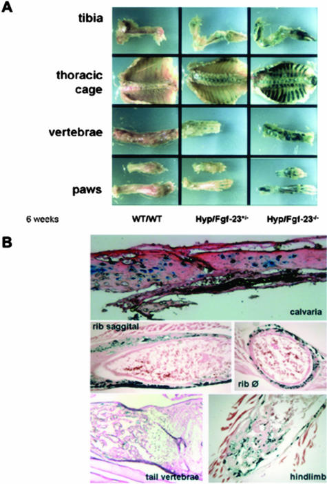
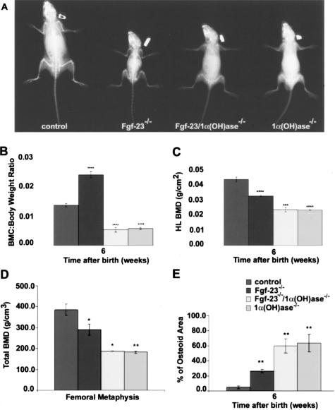
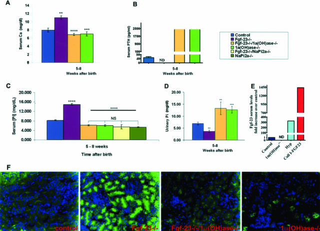
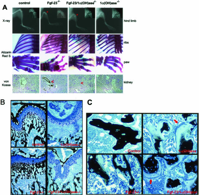
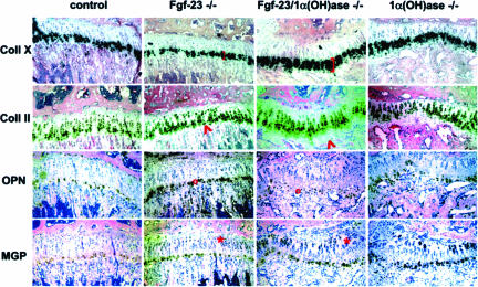
Fibroblast growth factor-23 (FGF-23) is one of the circulating phosphaturic factors associated with renal phosphate wasting. Fgf-23-/- animals show extremely high serum levels of phosphate and 1,25-dihydroxyvitamin D3, along with abnormal bone mineralization and soft tissue calcifications. To determine the role of vitamin D in mediating altered phosphate homeostasis and skeletogenesis in the Fgf-23-/- mice, we generated mice lacking both the Fgf-23 and 1alpha-hydroxylase genes (Fgf-23-/-/1alpha(OH)ase-/-). In the current study, we have identified the cellular source of Fgf-23 in adult mice. In addition, loss of vitamin D activities from Fgf-23-/- mice reverses the severe hyperphosphatemia to hypophosphatemia, attributable to increased urinary phosphate wasting in Fgf-23-/-/1alpha(OH)ase-/- mice, possibly as a consequence of decreased expression of NaPi2a. Ablation of vitamin D from Fgf-23-/- mice resulted in further reduction of total bone mineral content and bone mineral density and reversed ectopic calcification of skeleton and soft tissues, suggesting that abnormal mineral ion homeostasis and impaired skeletogenesis in Fgf-23-/- mice are mediated through enhanced vitamin D activities. In conclusion, using genetic manipulation studies, we have provided evidence for an in vivo inverse correlation between Fgf-23 and vitamin D activities and for the severe skeletal and soft tissue abnormalities of Fgf-23-/- mice being mediated through vitamin D.


Figures

Figure 1
Figure 1
Expression of Fgf-23 by β-galactosidase staining at 6 weeks. A: Shown are various skeletal elements including tibia, thoracic cage, vertebrae, and paws of wild-type (WT/WT) mouse, an Fgf-23 heterozygous mouse in a Hyp mouse background (Hyp+/−/Fgf-23+/−), and an Fgf-23 homozygous mutant mouse in a Hyp mouse background (Hyp+/−/Fgf-23−/−). Please note the difference in intensity of β-galactosidase staining in Hyp+/−/Fgf-23+/− versus Hyp+/−/Fgf-23−/− bones. B: Represented are frozen sections of stained Hyp+/−/Fgf-23−/− bones such as calvaria, ribs (saggital and transverse), tail vertebrae, and tibia/hindlimb; specific blue staining is only evident in osteocytes of intramembranous and endochondral formed bones. No staining is evident in osteoblasts or cartilaginous areas.
Figure 2
Figure 2
A: X-ray autoradiography of total skeletons of a control, Fgf-23−/−, Fgf-23−/−/1α(OH)ase−/−, and 1α(OH)ase−/− animal at 6 weeks. B–D: Total BMC (B, each value obtained for BMC was normalized to the body weight of the corresponding animal), and BMD (C and D) by PIXImus and pQCT of hindlimbs of control, Fgf-23−/−, Fgf-23−/−/1α(OH)ase−/−, and 1α(OH)ase−/− animals are shown. E: Quantitative histomorphometry on osteoid volume of control, Fgf-23−/−, Fgf-23−/−/1α(OH)ase−/−, and 1α(OH)ase−/− animals (statistical significance: *P < 0.05, **P < 0.01,***P < 0.001, ****P < 0.0001). There was no statistical significant difference between WT (Fgf-23+/+/1α(OH)ase+/+) and Fgf-23+/−/1α(OH)ase+/−, Fgf-23−/− and Fgf-23−/−/1α(OH)ase+/−, and Fgf-23−/−/1α(OH)ase−/− and 1α(OH)ase−/−/Fgf-23+/− animals, so data were combined.
Figure 3
Figure 3
Biochemical measurements of calcium, phosphate, PTH, and FGF-23 (A–E), and NaPi2a immunohistochemistry in various mouse mutants (F). Serum calcium (A) levels in control (n = 15), Fgf-23−/− (n = 6), Fgf-23−/−/1α(OH)ase−/− (n = 8), and 1α(OH)ase−/− (n = 11) animals; serum PTH (B) levels in control (n = 8), Fgf-23−/− (n = 4), Fgf-23−/−/1α(OH)ase−/− (n = 3), and 1α(OH)ase−/− (n = 9) animals; serum phosphate (C) levels in control (n = 22), Fgf-23−/− (n = 11), Fgf-23−/−/1α(OH)ase−/− (n = 7), 1α(OH)ase−/− (n = 14), Fgf-23−/−/NaPi2a−/− (n = 3), and NaPi2a−/− (n = 3) animals; urinary phosphate (D) control (n = 9), Fgf-23−/− (n = 5), Fgf-23−/−/1α(OH)ase−/− (n = 5), and 1α(OH)ase−/− (n = 9) animals; and serum Fgf-23 (E) levels in control (n = 8), 1α(OH)ase−/− (n = 5), Hyp (light blue, n = 1), and Coll I-FGF23 (red, n = 2) animals were measured in 5- to 12-week-old mice. Statistical significance: **P < 0.01, ***P < 0.001, ****P < 0.0001. Immunostaining of NaPi2a in the kidney sections prepared from wild-type, Fgf-23−/−, Fgf-23−/−/1α(OH)ase−/−, and 1α(OH)ase−/− animals using a polyclonal antibody (F). Please note that in contrast to the increased expression of NaPi2a in Fgf-23−/− mice, there is significantly less expression of NaPi2a protein in the Fgf-23−/−/1α(OH)ase−/− mice, similar to the expression in mice that lack the 1α(OH)ase gene [1α(OH)ase−/−].
Figure 4
Figure 4
A: Top: X-ray autoradiography of hindlimbs from a control, Fgf-23−/−, Fgf-23−/−/1α(OH)ase−/−, and 1α(OH)ase−/− mouse at 6 weeks. Asterisk depicts the ricketic widening of the growth plate in double-mutant versus Fgf-23−/− mice. Middle: Alizarin Red S-stained skeletal elements (ribs and paws) from each animal demonstrate loss of nodule formation in Fgf23−/−/1α(OH)ase−/− mutants. Bottom: Von Kossa staining of kidneys of each genotype. Red arrowheads point to the calcification of renal vessels of Fgf-23−/−, which is completely eliminated in Fgf-23−/−/1α(OH)ase−/− double mutants. B: Three-μm-thick undecalcified sections from 6-week-old control, Fgf-23−/−, Fgf-23−/−/1α(OH)ase−/−, and 1α(OH)ase−/− bones were stained with von Kossa/McNeal. Black staining represents mineralization. More mineral deposition is found in the area below the growth plate (methaphysis) in Fgf-23−/− mice. In contrast, large areas of unmineralized osteoid (light blue) are found in bones of Fgf-23−/−/1α(OH)ase−/− and 1α(OH)-ase−/− mice. C: Cancellous bone of 1α(OH)ase−/− and Fgf-23−/−/1α(OH)ase−/− compound mutants show hyperactive, cuboidal osteoblasts on top of extremely thick osteoid layers (red arrows). Osteoblasts in Fgf-23−/− mice appeared more flat, and osteoid seams were thinner. Original magnifications, ×20 (A, B).
Figure 5
Figure 5
In situ hybridization with riboprobes for collagen type X (Coll X), collagen type II (Coll II), osteopontin (OPN), and matrix gla protein (MGP) on sections from tibia of control, Fgf-23−/−, Fgf-23−/−/1α(OH)ase−/−, and 1α(OH)ase−/− at 6 weeks. Brackets depict the size of the zone of hypertrophic chondrocytes. Red arrowheads point to an area of hypertrophic chondrocytes that is only present in Fgf-23−/−/1α(OH)ase−/− and 1α(OH)ase−/− bones. Red circles show the decrease in OPN expression in the ricketic phenotype, and red asterisk depicts an expansion of MGP expression in the growth plate of Fgf-23−/−/1α(OH)ase−/− and 1α(OH)ase−/− bones.

References

    1. Bringhurst FR, Demay MB, Kronenberg HM. Hormones and disorders of mineral metabolism. Philadelphia: WB Saunders Co.,; Williams Textbook of Endocrinology. 1998:pp 1155–1210.
    1. Shimada T, Mizutani S, Muto T, Yoneya T, Hino R, Takeda S, Takeuchi Y, Fujita T, Fukumoto S, Yamashita T. Cloning and characterization of FGF23 as a causative factor of tumor-induced osteomalacia. Proc Natl Acad Sci USA. 2001;98:6500–6505. - PMC - PubMed
    1. White KE, Evans WE, O’Riordan JLH, Speer MC, Econs MJ, Lorenz-Depiereux B, Grabowski M, Mettinger T, Strom TM. Autosomal dominant hypophosphataemic rickets is associated with mutations in FGF23. The ADHR Consortium. Nat Genet. 2000;26:345–348. - PubMed
    1. Francis F, Henning S, Korn B, Reinhardt R, de Jong P, Poustka A, Lehrach H, Rowe PSN, Goulding JN, Summerfield T, Mountford R, Read AP, Popowska E, Pronicka E, Davies KE, O’Riordan JLH, Econs MJ, Nesbitt T, Drezner MK, Oudet C, Pannetier S, Hanauer A, Strom TM, Meindl A, Lorenz B, Cagnoli M, Mohnike KL, Murken J, Meitinger T. A gene (PEX) with homologies to endopeptidases is mutated in patients with X-linked hypophosphatemic rickets. The HPY Consortium. Nat Genet. 1995;11:130–136. - PubMed
    1. Shimada T, Urakawa I, Yamazaki Y, Hasegawa H, Hino R, Yoneya T, Takeuchi Y, Fujita T, Fukumoto S, Yamashita T. FGF-23 transgenic mice demonstrate hypophosphatemic rickets with reduced expression of sodium phosphate cotransporter type IIa. Biochem Biophys Res Commun. 2004;314:409–414. - PubMed

Publication types

MeSH terms