. 2006 Dec;169(6):2199-208.
doi: 10.2353/ajpath.2006.060171.

Suppressor of cytokine signaling-3 antagonizes cAMP effects on proliferation and apoptosis and is expressed in human prostate cancer

Affiliations

Suppressor of cytokine signaling-3 antagonizes cAMP effects on proliferation and apoptosis and is expressed in human prostate cancer

Ilaria Bellezza et al. Am J Pathol. 2006 Dec.

Abstract

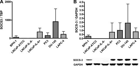
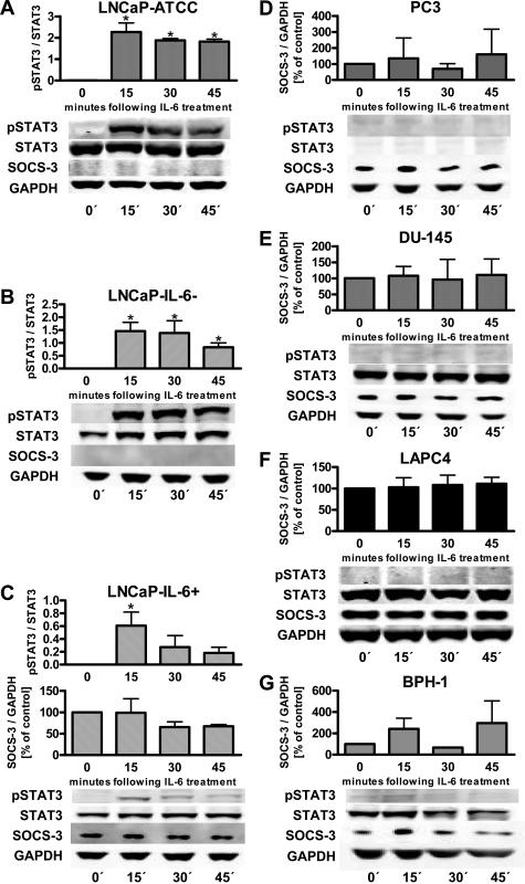
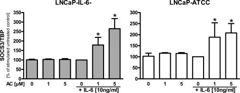
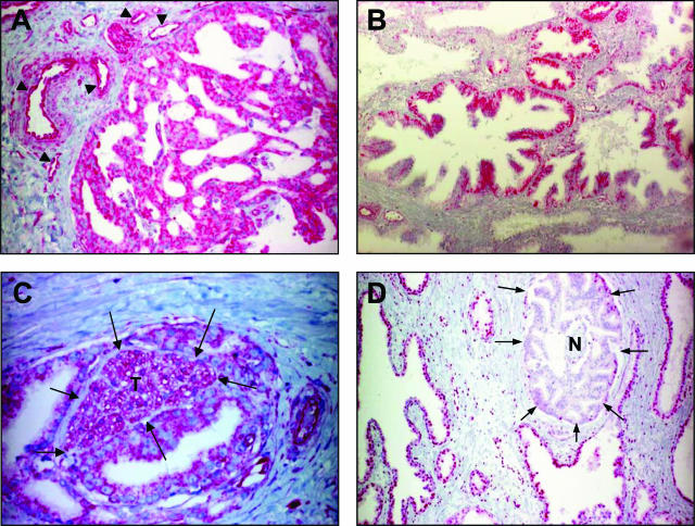
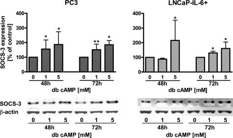
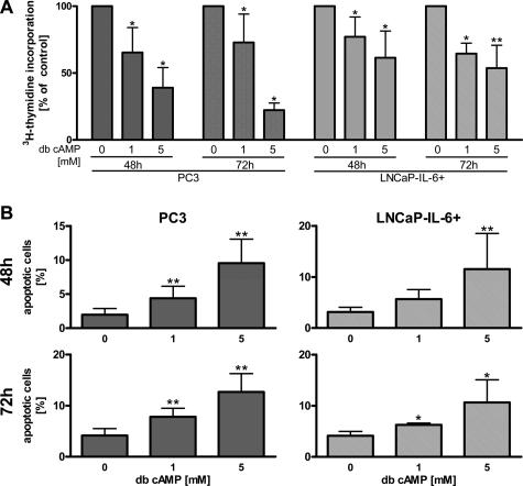
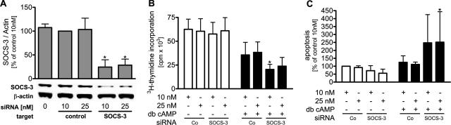
Interleukin-6, levels of which are elevated in prostate cancer, activates different signal transduction pathways including that of Janus kinases/signal transducer and activator of transcription (STAT)3. However, phosphorylation of STAT3 has been reported to be associated with either stimulatory or inhibitory effects on cellular proliferation. To better understand the mechanisms of STAT3 regulation in benign and malignant prostate, we have investigated the role of suppressor of cytokine signaling (SOCS)-3. Cell lines that did not express phosphorylated STAT3 were found to be SOCS-3-positive. SOCS-3 was re-expressed in LNCaP cells after treatment with a demethylating agent. SOCS-3 immunohistochemistry revealed a negative or weak reaction in benign areas, whereas its expression was detected in tumor tissue. To investigate the involvement of SOCS-3 in regulation of cellular events, we incubated cancer cells with a cAMP derivative. This treatment yielded higher SOCS-3 levels, reduced [3H]thymidine incorporation, and increased percentage of apoptotic cells. However, down-regulation of SOCS-3 by a short interfering RNA approach resulted in inhibition of proliferation and an increased apoptotic rate. Collectively, our results show that SOCS-3 antagonizes regulation of cellular events by cAMP and is expressed in human prostate cancer.


Figures

Figure 1
Figure 1
Basal expression of SOCS-3 mRNA (A) and protein (B) in benign and malignant prostate cells. SOCS-3 was measured by real-time PCR and Western blot, respectively. The values were normalized according to the housekeeping genes TBP and GAPDH, respectively (n = 3, mean values ± SD).
Figure 2
Figure 2
Correlation between expression of SOCS-3 and expression and phosphorylation of STAT3 in prostate cells. SOCS-3, pSTAT3, STAT3, and GAPDH were determined by Western blotting after treatment with IL-6. Densitometric analysis and results of representative experiments are shown (n = 3, mean values ± SD, *P < 0.05, IL-6-treated versus untreated cells, Mann-Whitney U-test).
Figure 3
Figure 3
Re-expression of SOCS-3 after treatment with the demethylating agent 5-aza-2′-cytidine (AC). LNCaP-IL-6 and LNCaP cells were treated with increasing concentrations of 5-aza-2′-cytidine for 4 days followed by a 1-hour incubation with 10 ng/ml IL-6. The cells were then harvested, and RNA was isolated and transcribed in cDNA. qRT-PCR was then performed. The results are expressed in relation to values measured in untreated cells and represent mean values ± SD from at least three independent experiments (*P < 0.05, Mann-Whitney U-test).
Figure 4
Figure 4
Immunohistochemical expression of SOCS-3 (A–C) and pSTAT3 (D) in benign and malignant prostate tissue. Specimens obtained from patients were stained with the anti-SOCS-3 antibody diluted 1:100 or anti-pSTAT3 antibody diluted 1:20. Expression of SOCS-3 in endothelium (A, arrowheads) was regarded as a positive control. SOCS-3 expression is detected in Gleason pattern 3 prostate cancer specimen (A), high-grade prostate intraepithelial neoplasia (B), and tumor (T, arrows) that invades perineural space (C). pSTAT3 immunostaining is shown in benign tissue but not in tumor cells (D) (arrows, tumor; N, nerve). Original magnifications: ×20 (A, B); ×40 (C); ×10 (D).
Figure 5
Figure 5
Expression of SOCS-3 after treatment with the cAMP analogue db cAMP. PC3 and LNCaP-IL-6+ cell lines were treated with increasing concentrations of db cAMP for 48 and 72 hours before determination of SOCS-3 protein levels. Results of densitometric analysis and representative Western blots are shown (n ≥ 4, mean values ± SD, *P < 0.05, db cAMP-treated versus untreated cells, Mann-Whitney U-test).
Figure 6
Figure 6
Regulation of cellular proliferation (A) and apoptosis (B) in PC3 and LNCaP-IL-6+ cells. The two cell lines were treated with db cAMP (1 and 5 mmol/L) for 48 and 72 hours. Cellular proliferation was assessed by measurement of [3H]thymidine incorporation, and the percentage of apoptotic cells was determined by flow cytometry (n ≥ 4, mean values ± SD, *P < 0.05, **P < 0.01, db cAMP-treated versus untreated cells, Mann-Whitney U-test). In A, the percentage of untreated cells was set at 100.
Figure 7
Figure 7
A: Expression of SOCS-3 after transfection of PC3 cells with control or SOCS-3 siRNA (10 or 25 nmol/L). Cells were transfected using Fugene 6, and protein expression was analyzed by Western blot. Mock transfection (first column) was performed as an additional control. Cellular proliferation was assessed by [3H]thymidine incorporation (B) and flow cytometric analysis of sub-G1 peak cells was performed to measure apoptosis (C). Mann-Whitney U-test was used (n ≥ 4, mean values ± SD, control siRNA- versus SOCS-3 siRNA-treated cells in the presence of 5 mmol/L db cAMP, *P < 0.05).

References

    1. Giri D, Ozen M, Ittmann M. Interleukin-6 is an autocrine growth factor in human prostate cancer. Am J Pathol. 2001;159:2159–2165. - PMC - PubMed
    1. Twillie DA, Eisenberger MA, Carducci MA, Hseih W-S, Kim WY, Simons JW. Interleukin-6: a candidate mediator of human prostate cancer morbidity. Urology. 1995;45:542–549. - PubMed
    1. Liu XH, Kirschenbaum A, Lu M, Yao S, Klausner A, Preston C, Holland JF, Levine AC. Prostaglandin E(2) stimulates prostatic intraepithelial neoplasia cell growth through activation of the interleukin-6/GP130/STAT-3 signaling pathway. Biochem Biophys Res Commun. 2002;290:249–255. - PubMed
    1. Chung TD, Yu JJ, Kong TA, Spiotto MT, Lin JM. Interleukin-6 activates phosphatidylinositol-3 kinase, which inhibits apoptosis in human prostate cancer cell lines. Prostate. 2000;42:1–7. - PubMed
    1. Mora LB, Buettner R, Seigne J, Diaz J, Ahmad N, Garcia R, Bowman T, Falcone R, Fairclough R, Cantor A, Muro-Cacho C, Livingston S, Karras J, Pow-Sang J, Jove R. Constitutive activation of Stat3 in human prostate tumors and cell lines: direct inhibition of Stat3 signaling induces apoptosis of prostate cancer cells. Cancer Res. 2002;62:6659–6666. - PubMed

Publication types

MeSH terms