Review
. 2007;29(1-2):14-27.
doi: 10.1159/000096208.

Ectopic granule cells of the rat dentate gyrus

Affiliations
Review

Ectopic granule cells of the rat dentate gyrus

Helen Scharfman et al. Dev Neurosci. 2007.

Abstract

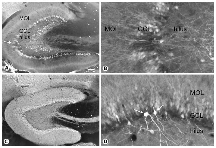
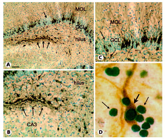
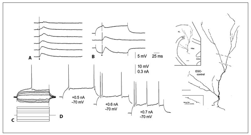
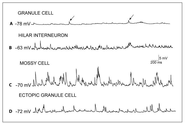
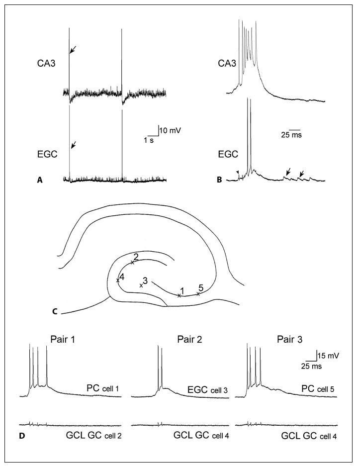
Granule cells of the mammalian dentate gyrus normally form a discrete layer, and virtually all granule cells migrate to this location. Exceptional granule cells that are positioned incorrectly, in 'ectopic' locations, are rare. Although the characteristics of such ectopic granule cells appear similar in many respects to granule cells located in the granule cell layer, their rare occurrence has limited a full evaluation of their structure and function. More information about ectopic granule cells has been obtained by studying those that develop after experimental manipulations that increase their number. For example, after severe seizures, the number of ectopic granule cells located in the hilus increases dramatically. These experimentally-induced ectopic granule cells may not be equivalent to normal ectopic granule cells necessarily, but the vastly increased numbers have allowed much more information to be obtained. Remarkably, the granule cells that are positioned ectopically develop intrinsic properties and an axonal projection that are similar to granule cells that are located normally, i.e., in the granule cell layer. However, dendritic structure and synaptic structure/function appear to differ. These studies have provided new insight into a rare type of granule cell in the dentate gyrus, and the plastic characteristics of dentate granule cells that appear to depend on the location of the cell body.


Figures

Fig. 1
Fig. 1
Calbindin expression in a control and epileptic rat. A Calbindin expression in a rat that had status epilepticus and recurrent seizures shows irregular loss of calbindin expression in the granule cell layer (GCL) and numerous ectopically located calbindin-immunoreactive cells throughout the deep hilus. MOL = Molecular layer. Calibration = 100 μm. For methods used for calbindin immunocytochemistry, see Scharfman et al. [2000, . B Higher magnification of the area in A marked by the arrow. Same orientation as in A. Calibration (in A) = 50 μm. C Calbindin expression in a saline-treated rat that was of a similar age at the time of euthanasia as the rat in A. Note the even, homogeneous expression of calbindin throughout the granule cell layer and the lamina containing granule cell dendrites and the mossy fiber pathway. However, calbindin-immunoreactive cells are not present in the hilus. Same calibration as in A. D Example of unusual calbindin immunoreactivity in a pilocarpine-treated rat with chronic seizures. Dense reactivity is apparent in nongranule cells (arrows) relative to granule cells, suggesting seizure-induced up-regulation of calbindin expression in nongranule cells at the same time as seizure-induced downregulation of calbindin in granule cells. Same calibration as in B. MOL = Molecular layer; GCL = granule cell layer.
Fig. 2
Fig. 2
Calbindin and PROX1 expression in the epileptic rat. MOL = Molecular layer; GCL = granule cell layer. A Calbindin and cresyl violet staining of a section through the septal pole of a pilocarpine-treated rat with recurrent seizures illustrates irregular loss of calbindin expression despite persistence of apparently normal granule cell cresyl violet staining (arrow) and clusters of calbindin-immunoreactive cells at the border of the hilus and CA3 region (triple arrows). Calibration = 100 μm. B Higher magnification of the clusters of hilar calbindin cells shown in A. Calibration (in A) = 50 μm. C Higher magnification of the irregular loss of calbindin in the granule cell layer (arrow in A). Calibration as for B. D Double-labeling of sections from the hilus of a pilocarpine-treated rat with recurrent seizures demonstrates that numerous PROX1-immunoreactive nuclei are present (small arrows), but only one is double-labeled with calbindin (orange, large arrow), suggesting that PROX1 is the more reliable marker of mature granule cells than calbindin in rats with chronic seizures. Calibration (in A) = 5 μm.
Fig. 3
Fig. 3
Characteristics of ectopic hilar granule cells in the normal rat. A Synaptic responses to outer molecular layer stimulation of the cell shown on the far right, an ectopic granule cell located in the deep hilus of a normal adult rat. B Synaptic responses at different holding potentials illustrate the common synaptic response of a granule cell to molecular layer stimulation in vitro. C, D Intracellular current injection evoked responses in the ectopic cell that were similar to granule cells in the granule cell layer. For further details, see Scharfman et al. [2003]. Reprinted from Neuroscience. EGC = Ectopic granule cell.
Fig. 4
Fig. 4
Comparison of spontaneous synaptic activity in neurons recorded intracellularly in hippocampal slices of rats with spontaneous recurrent seizures. A Representative recording from a granule cell located in the granule cell layer at a hyperpolarized potential shows spontaneous depolarizations (arrows), which are likely to be IPSPs, because they reversed polarity at approximately −70 mV (not shown), typical of GABAergic IPSPs under these recording conditions. B Example of spontaneous activity in a GABAergic interneuron located in the hilar region. C Spontaneous activity in a mossy cell demonstrates the typical high frequency and large amplitude of spontaneous depolarizations recorded in these cells. D A recording from an ectopic granule cell shows a higher frequency and amplitude of spontaneous events relative to granule cells located in the normal position (compare to A). The spontaneous potentials are likely to be a mixture of IPSPs and EPSPs, because of their reversal potentials (not shown). Thus, some events reverse at −70 mV, like IPSPs mediated by Cl, and the others reverse at much more depolarized potentials, like EPSPs.
Fig. 5
Fig. 5
Abnormal synchronized bursts of ectopic hilar granule cells with CA3 neurons, recorded in hippocampal slices from rats with spontaneous recurrent seizures. A A continuous recording from a CA3 pyramidal cell (top, CA3) and an ectopic granule cell (bottom, EGC) in a slice from a rat that had spontaneous recurrent seizures. There are two spontaneous bursts of action potentials that are synchronized. The arrows point to one of the synchronized events. B One of the synchronous events in A is shown with higher temporal resolution. The arrowhead marks the capacitative artifact of the CA3 pyramidal cell’s first action potential in the recording of the ectopic granule cell. It shows that the first action potential of CA3 occurs at the onset of the depolarization in the ectopic granule cell. The arrows denote spontaneous synaptic depolarizations. C The schematic illustrates recording positions for D. D Three consecutively recorded pairs of simultaneous intracellular recordings are shown. Recordings were from the same slice, from a rat that had status epilepticus and spontaneous, recurrent seizures. Left: the first pair of cells to be recorded simultaneously was a CA3 pyramidal cell (top, PC) and a granule cell (GC) of the granule cell layer (bottom, GCL). The CA3 recording shows the intracellular correlate of the population burst discharge: a large depolarization with several action potentials at its peak. There was no activity in the simultaneously recorded granule cell when the CA3 pyramidal cell had the spontaneous depolarization and discharge of action potentials. This was true at the potential of the recording, −70 mV, and other membrane potentials between −50 and −80 mV (data not shown). There are four small deflections in the granule cell recording that do not reflect activity, but are the capacitative artifacts of the action potentials in the simultaneous CA3 pyramidal cell recording. Center: the second pair of cells included an ectopic hilar granule cell (EGC) and a second granule cell (GC) from the granule cell layer (GCL), which were both impaled after the first two cells shown at left. The neuron from the granule cell layer was silent when there was a burst discharge that occurred in the ectopic granule cell. Right: another CA3 pyramidal cell (PC) was impaled subsequent to the ectopic granule cell shown in the center, while maintaining the impalement of the granule cell (GC) in the granule cell layer (GCL) shown in the center. During the spontaneous burst discharge of the CA3 neuron, the granule cell demonstrated little activity. These recordings demonstrate that during the spontaneous discharges of CA3 and hilar ectopic neurons, there is no evidence of synchronized burst discharges in the granule cells that are located in the normal position, the granule cell layer. Note, however, that there can be subthreshold depolarizations that follow the burst after 5–25 ms, suggesting that recurrent excitatory circuits may activate granule cells after the CA3-ectopic bursts occur. For further discussion, see Scharfman et al. [2000, , and Scharfman [2004].

Similar articles

Cited by

References

    1. Åberg M, Åberg ND, Hedbacker H, Oscarsson J, Eriksson PS. Peripheral infusion of IGF-I selectively induces neurogenesis in the adult rat hippocampus. J Neurosci. 2000;20:2896–2903. - PMC - PubMed
    1. Acsady L, Katona I, Martinez-Guijarro FJ, Buzsáki G, Freund TF. Unusual target selectivity of perisomatic inhibitory cells in the hilar region of the rat hippocampus. J Neurosci. 2000;20:6907–6919. - PMC - PubMed
    1. Amaral DG. A Golgi study of cell types in the hilar region of the hippocampus in the rat. J Comp Neurol. 1978;182:851–914. - PubMed
    1. Amaral DG. Synaptic extensions from the mossy fibers of the fascia dentata. Anat Embryol. 1979;155:241–251. - PubMed
    1. Amaral DG, Woodward DJ. A hippocampal interneuron observed in the inferior region. Brain Res. 1977;124:225–236. - PubMed

Publication types

MeSH terms