. 2007 Feb 9;144(3):991-1003.
doi: 10.1016/j.neuroscience.2006.09.064. Epub 2006 Dec 5.

Depletion of reduced glutathione enhances motor neuron degeneration in vitro and in vivo

Affiliations

Depletion of reduced glutathione enhances motor neuron degeneration in vitro and in vivo

L Chi et al. Neuroscience. .

Abstract

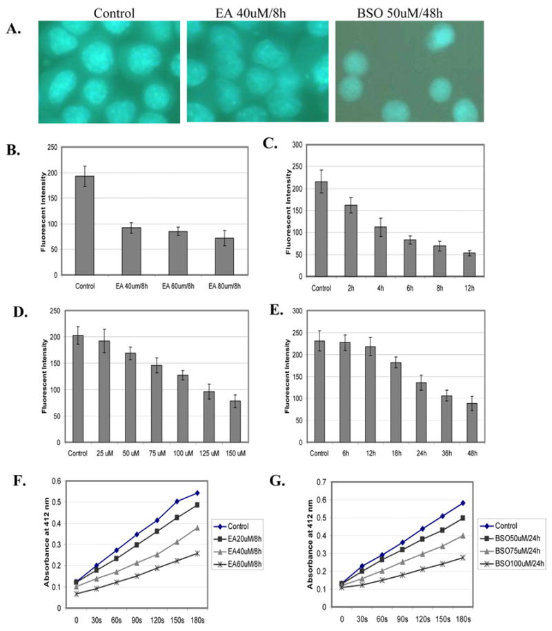
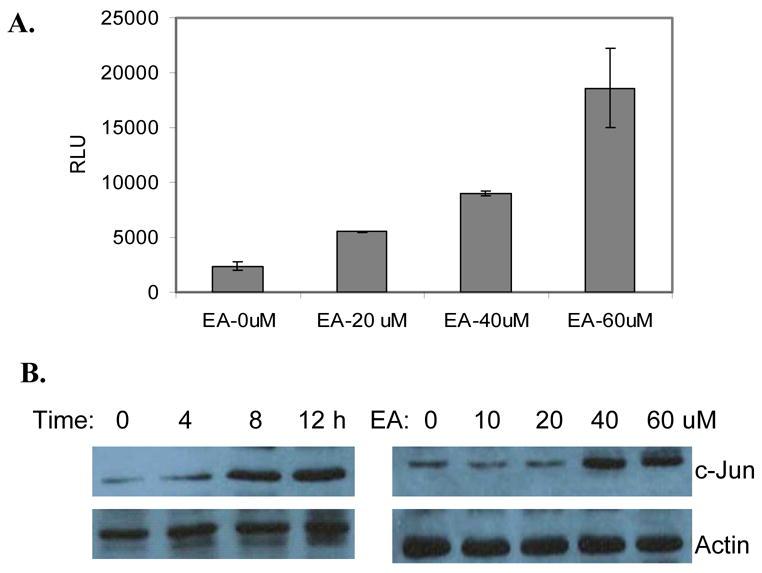
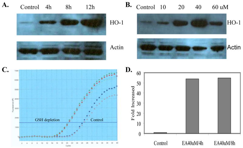
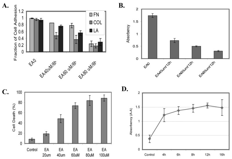
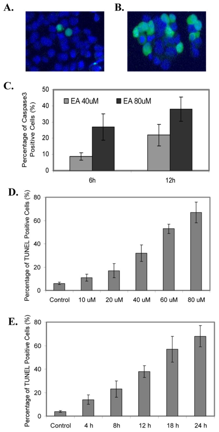
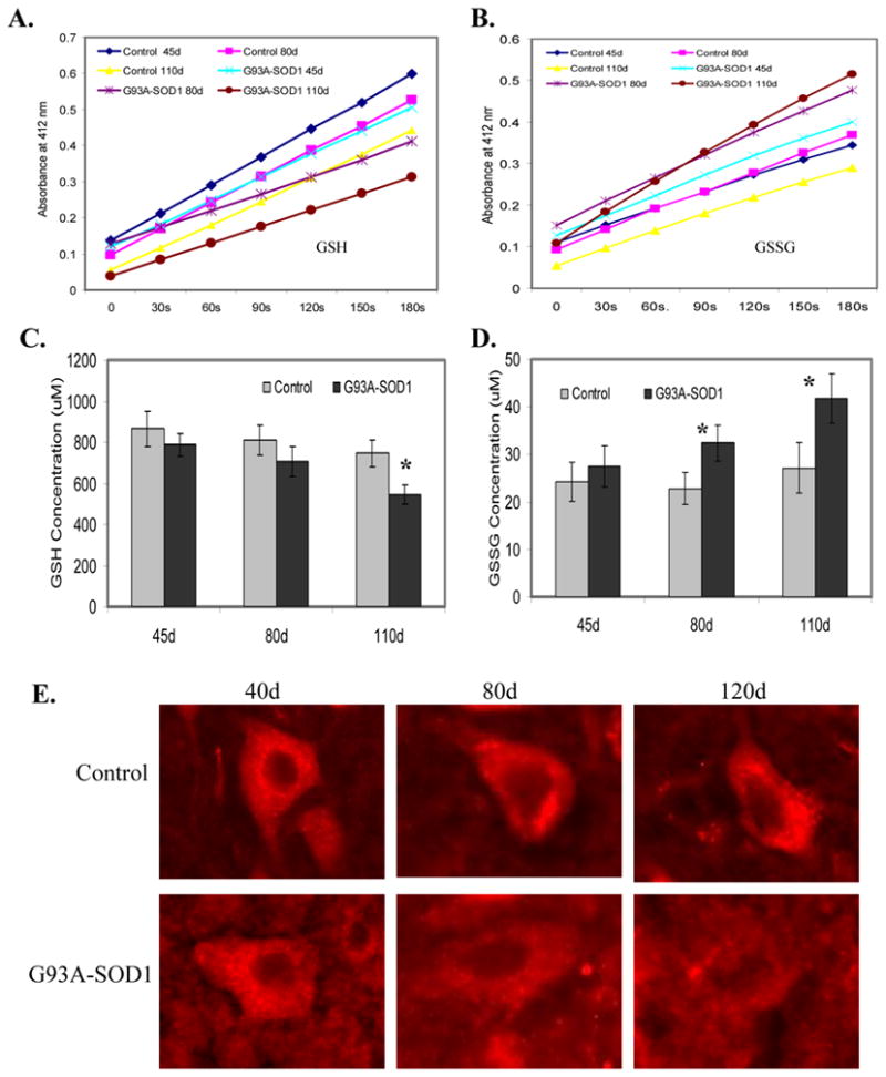
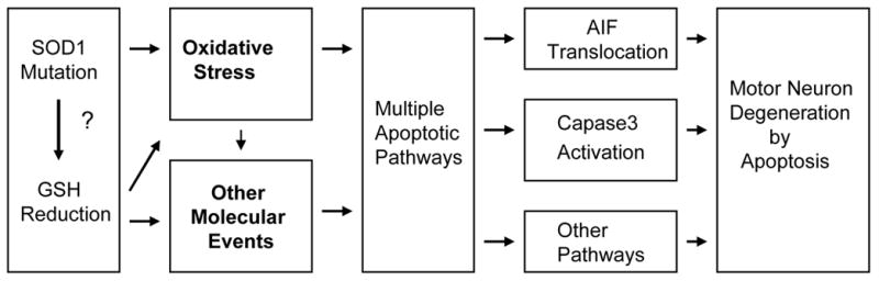
The mechanism of selective and age-dependent motor neuron degeneration in human amyotrophic lateral sclerosis (ALS) has not been defined and the role of glutathione (GSH) in association with motor neuron death remains largely unknown. A motor neuron-like cell culture system and a transgenic mouse model were used to study the effect of cellular GSH alteration on motor neuron cell death. Exposure of NSC34 motor neuron-like cells to ethacrynic acid (EA) or l-buthionine sulfoximine (BSO) dramatically reduced the cellular GSH levels, and was accompanied by increased production of reactive oxygen species (ROS) measured by the dichlorofluorescin (DCF) fluorescent oxidation assay. In addition, GSH depletion enhanced oxidative stress markers, AP-1 transcriptional activation, c-Jun, c-Fos and heme oxygenase-1 (HO-1) expression in NSC34 cells analyzed by a luciferase reporter, Western blotting and quantitative PCR assays respectively. Furthermore, depletion of GSH decreased mitochondrial function, facilitated apoptosis inducing factor (AIF) translocation, cytochrome c release, and caspase 3 activation, and consequently led to motor neuron-like cell apoptosis. In an ALS-like transgenic mouse model overexpressing mutant G93A-Cu, Zn-superoxide dismutase (SOD1) gene, we showed that the reduction of GSH in the spinal cord and motor neuron cells is correlated with AIF translocation, caspase 3 activation, and motor neuron degeneration during ALS-like disease onset and progression. Taken together, the in vitro and in vivo data presented in the current report demonstrated that decreased GSH promotes multiple apoptotic pathways contributing, at least partially, to motor neuron degeneration in ALS.


Figures

Figure 1
Figure 1. Chemical depletion of GSH production and biochemical inhibition of GSH synthesis in NSC34 motor neuron-like cells
A. Representative images of control, EA and BSO treated cells demonstrating reduction of GSH analyzed by monobromobimane (MBM) assay. B. GSH depletion by EA for 8h in NSC34 cells causes a dose-dependent decrease of intracellular GSH detected with MBM assay. C. GSH depletion by EA at 60 μM in NSC34 cells causes a time-dependent decrease of intracellular GSH detected with MBM assay. D. GSH synthesis inhibition by BSO for 24h in NSC34 cells causes a dose-dependent decrease of intracellular GSH detected with MBM assay. E. GSH synthesis inhibition by BSO at 75 μM in NSC34 cells causes a time-dependent decrease of intracellular GSH detected with MBM assay. F. GSH depletion by EA in NSC34 cells causes a dose-dependent decrease of intracellular GSH detected with kinetic assay (refer to the detailed assay in the Experimental Method Section). G. GSH synthesis inhibition by BSO in NSC34 cells causes a dose-dependent decrease of intracellular GSH detected with kinetic assay (refer to the detailed assay in the Experimental Method Section). All experiments were carried out with triplicate and three independent repeats.
Figure 2
Figure 2. GSH depletion enhances reactive oxygen species (ROS) production in NSC34 cells
A. GSH depletion by EA in NSC34 cells causes a dose-dependent increase of intracellular ROS production detected with DCF assay. B. GSH synthesis inhibition by BSO in NSC34 cells causes a dose-dependent increase of intracellular ROS production detected with DCF assay. All experiments were carried out with tetra-plicate and three independent repeats.
Figure 3
Figure 3. GSH depletion by EA increases early oxidative response gene expression
A. GSH depletion by EA in NSC34 cells enhances AP-1 transcriptional activation detected by a luciferase reporter assay. B. GSH depletion by EA in NSC34 cells increases a dose and time-dependent c-Jun expression detected by western blotting analysis.
Figure 4
Figure 4. GSH depletion by EA increases secondary oxidative response gene expression
A. GSH depletion by EA at 40 μM in NSC34 cells dramatically increases a time-dependent HO-1 expression detected by western blotting analysis. B. GSH depletion by EA for 12h in NSC34 cells increases a dose-dependent HO-1 expression detected by western blotting analysis. C. GSH depletion by EA at 40 μM for 4h and 8h in NSC34 cells dramatically increases HO-1 expression detected by quantitative real-time PCR analysis. D. Fold-increase of HO-1 expression in EA treated cells compared to vehicle control detected by quantitative real-time PCR analysis (n=3).
Figure 5
Figure 5. GSH depletion by EA increases NSC34 cell death
A. GSH depletion by EA in NSC34 cells decreases cell adhesion to fibronectin (FN), collagen (COL) and laminin (LA) in a dose-dependent manner. B. GSH depletion by EA in NSC34 cells increases mitochondrial dysfunction in a dose-dependent manner analyzed by MTT assay. C. GSH depletion by EA in NSC34 cells increases cell death in a dose-dependent manner detected by trypan blue exclusion assay. D. GSH depletion by EA in NSC34 cells increases cell apoptosis in a time-dependent manner detected by histone protein release assay.
Figure 6
Figure 6. GSH depletion by EA promotes redistribution of pro-apoptotic factors in NSC34 cells
A. GSH depletion by EA in NSC34 cells enhances AIF nuclear translocation from mitochondria detected by western blotting analysis coupled with mitochondrial and nuclear fractionation. B. GSH depletion by EA in NSC34 cells promotes cytochrome c release to cytosol from mitochondria detected by western blotting analysis coupled with mitochondrial and cytosolic fractionation. C. GSH depletion by EA in NSC34 cells enhances AIF nuclear translocation from mitochondria detected by immunohistochemical staining. D. GSH depletion by EA in NSC34 cells promotes cytochrome c release from mitochondria detected by immunohistochemical staining.
Figure 7
Figure 7. GSH depletion by EA promotes apoptosis in NSC34 cells
A. Vehicle control treated NSC34 cells are immunostained with active caspase 3, showing that most of cells are negative in active caspase 3 staining. B. GSH depletion by EA in NSC34 cells enhances caspase 3 activation and increases nuclear translocation detected by immunohistochemical staining. C. Percentage of active caspase 3 positive cells in EA treated NSC34 cells (n=3). D. GSH depletion by EA in NSC34 cells promotes a dose-dependent cell apoptosis detected by TUNEL assay. E. GSH depletion by EA in NSC34 cells promotes a time-dependent cell apoptosis detected by TUNEL assay.
Figure 8
Figure 8. GSH levels in mutant G93A-SOD1 transgenic mice at disease free (45 days of age), disease onset (80 days of age) and disease progression (110 days of age) compared to the age-matched normal mouse controls
Spinal cord lumbar region from mutant G93A-SOD1 transgenic mice and age-matched control mice were processed to determine the levels of GSH or GSSG. A. Representative kinetic assay of GSH (refer to the detailed assay in the Experimental Method Section) in the spinal cord lumbar region of mutant G93A-SOD1 transgenic mice compared to the age-matched normal mouse controls. B. Representative kinetic assay of GSSG (refer to the detailed assay in the Experimental Method Section) in the spinal cord lumbar region of mutant G93A-SOD1 transgenic mice compared to the age-matched normal mouse controls. C. The relative GSH levels in the spinal cord lumbar region of mutant G93A-SOD1 transgenic mice compared to the age-matched normal mouse controls. D. The relative GSSG levels in the spinal cord lumbar region of mutant G93A-SOD1 transgenic mice compared to the age-matched normal mouse controls. E. GSH levels in motor neuron cells of mutant G93A-SOD1 transgenic mice compared to the age-matched normal mouse controls.
Figure 9
Figure 9. Redistribution of AIF and active caspase 3 in motor neuron cells of ALS-like mice
Mutant G93A-SOD1 transgenic mice at disease progression stages (110 days of age) were processed to analyze the distribution of AIF and active caspase 3 in the spinal cord motor neurons. Compared with age-matched normal controls, AIF and active caspase 3 were translocated to the nuclei in the mutant G93A-SOD1 spinal cord motor neurons.
Figure 10
Figure 10
The potential signal transduction pathways of GSH depletion-mediated motor neuron cell apoptosis.

Similar articles

Cited by

References

    1. Adams JD, Jr, Klaidman LK, Odunze IN, Shen HC, Miller CA. Alzheimer's and Parkinson's disease. Brain levels of glutathione, glutathione disulfide, and vitamin E. Mol Chem Neuropathol. 1991;14:213–226. - PubMed
    1. Bains JS, Shaw CA. Neurodegenerative disorders in humans: the role of glutathione in oxidative stress-mediated neuronal death. Brain Res Brain Res Rev. 1997;25:335–358. - PubMed
    1. Bharath S, Hsu M, Kaur D, Rajagopalan S, Andersen JK. Glutathione, iron and Parkinson's disease. Biochem Pharmacol. 2002;64:1037–1048. - PubMed
    1. Bishop A, Marquis JC, Cashman NR, Demple B. Adaptive resistance to nitric oxide in motor neurons. Free Radic Biol Med. 1999;26:978–986. - PubMed
    1. Bogdanov MB, Ramos LE, Xu Z, Beal MF. Elevated "hydroxyl radical" generation in vivo in an animal model of amyotrophic lateral sclerosis. J Neurochem. 1998;71:1321–1324. - PubMed

Publication types

MeSH terms