

A novel subpopulation of 5-HT type 3A receptor subunit immunoreactive interneurons in the rat basolateral amygdala
- PMID: 17150309
- PMCID: PMC1828605
- DOI: 10.1016/j.neuroscience.2006.10.044
A novel subpopulation of 5-HT type 3A receptor subunit immunoreactive interneurons in the rat basolateral amygdala
Abstract
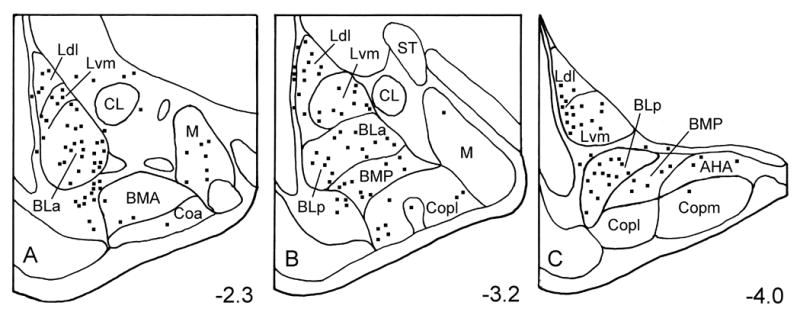
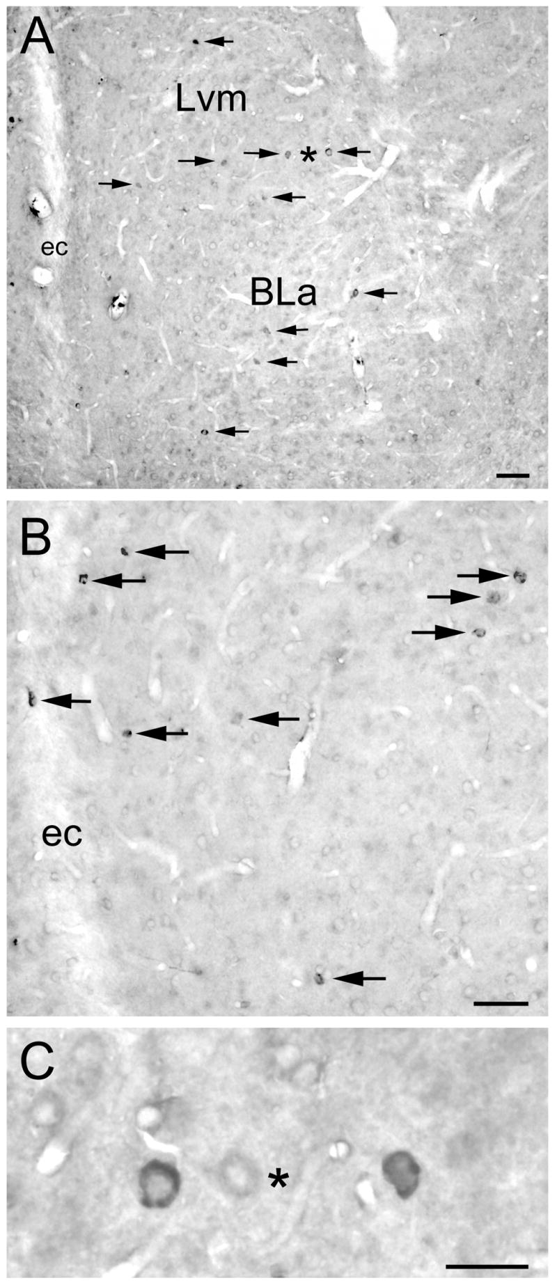
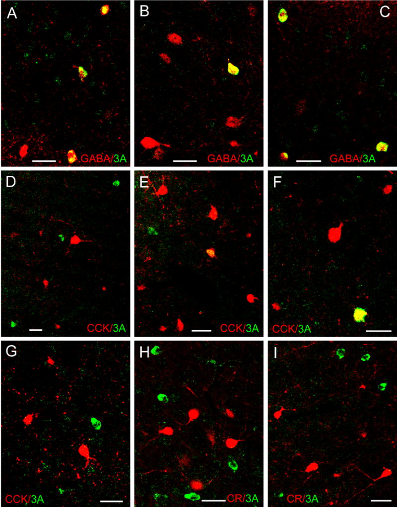
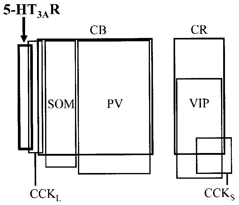
The amygdalar basolateral nuclear complex (BLC) has very high levels of the 5-HT type 3 receptor (5-HT(3)R). Previous studies have reported that 5-HT(3)R protein in the BLC is expressed in interneurons and that 5-HT(3)R mRNA is coexpressed with GABA and certain neuropeptides or calcium-binding proteins in these cells. However, there have been no detailed descriptions of the distribution of 5-HT(3)R+ neurons in the rat amygdala, and no quantitative studies of overlap of neurons expressing 5-HT(3)R protein with distinct interneuronal subpopulations in the BLC. The present investigation employed dual-labeling immunohistochemistry using antibodies to the 5-HT-3A receptor subunit (5-HT(3A)R) and specific interneuronal markers to address these questions. These studies revealed that there was a moderate density of nonpyramidal 5-HT(3A)R+ neurons in the BLC at all levels of the amygdala. In addition, immunostained cells were also seen in anterior portions of the cortical and medial nuclei. Although virtually all 5-HT(3A)R+ neurons in the BLC were GABA+, very few expressed neuropeptide or calcium-binding protein markers for individual subpopulations. The main interneuronal marker expressed by 5-HT(3A)R+ neurons was cholecystokinin (CCK), but only 8-16% of 5-HT(3)R+ neurons in the BLC, depending on the nucleus, were CCK+. Most of these CCK+/5-HT(3A)R+ double-labeled neurons appeared to belong to the subpopulation of large type L CCK+ interneurons. Very few 5-HT(3A)R+ neurons expressed calretinin, vasoactive intestinal peptide, or parvalbumin, and none expressed somatostatin or calbindin. Thus, the great majority of neurons expressing 5-HT(3A)R protein appear to constitute a previously unrecognized subpopulation of GABAergic interneurons in the BLC.
Figures
Similar articles
-
Differential expression of Kv3.1b and Kv3.2 potassium channel subunits in interneurons of the basolateral amygdala.Neuroscience. 2006;138(2):537-47. doi: 10.1016/j.neuroscience.2005.11.047. Epub 2006 Jan 18. Neuroscience. 2006. PMID: 16413129
-
Serotonin-immunoreactive axon terminals innervate pyramidal cells and interneurons in the rat basolateral amygdala.J Comp Neurol. 2007 Nov 20;505(3):314-35. doi: 10.1002/cne.21486. J Comp Neurol. 2007. PMID: 17879281
-
Immunohistochemical characterization of somatostatin containing interneurons in the rat basolateral amygdala.Brain Res. 2002 Jul 12;943(2):237-44. doi: 10.1016/s0006-8993(02)02650-1. Brain Res. 2002. PMID: 12101046
-
Neurochemical correlates of cortical GABAergic deficits in schizophrenia: selective losses of calcium binding protein immunoreactivity.Brain Res Bull. 2001 Jul 15;55(5):579-84. doi: 10.1016/s0361-9230(01)00526-3. Brain Res Bull. 2001. PMID: 11576754 Review.
-
Functional anatomy of 5-HT2A receptors in the amygdala and hippocampal complex: relevance to memory functions.Exp Brain Res. 2013 Oct;230(4):427-39. doi: 10.1007/s00221-013-3512-6. Epub 2013 Apr 17. Exp Brain Res. 2013. PMID: 23591691 Review.
Cited by
-
Building a 5-HT3A Receptor Expression Map in the Mouse Brain.Sci Rep. 2017 Mar 9;7:42884. doi: 10.1038/srep42884. Sci Rep. 2017. PMID: 28276429 Free PMC article.
-
Serotonin, Amygdala and Fear: Assembling the Puzzle.Front Neural Circuits. 2016 Apr 5;10:24. doi: 10.3389/fncir.2016.00024. eCollection 2016. Front Neural Circuits. 2016. PMID: 27092057 Free PMC article. Review.
-
Input-specific contributions to valence processing in the amygdala.Learn Mem. 2016 Sep 15;23(10):534-43. doi: 10.1101/lm.037887.114. Print 2016 Oct. Learn Mem. 2016. PMID: 27634144 Free PMC article. Review.
-
Mechanisms of inhibition within the telencephalon: "where the wild things are".Annu Rev Neurosci. 2011;34:535-67. doi: 10.1146/annurev-neuro-061010-113717. Annu Rev Neurosci. 2011. PMID: 21469958 Free PMC article. Review.
-
The Basolateral Amygdala Sends a Mixed (GABAergic and Glutamatergic) Projection to the Mediodorsal Thalamic Nucleus.J Neurosci. 2023 Mar 22;43(12):2104-2115. doi: 10.1523/JNEUROSCI.1924-22.2022. Epub 2023 Feb 14. J Neurosci. 2023. PMID: 36788026 Free PMC article.
References
-
- Abi-Dargham A, Laruelle M, Wong DT, Robertson DW, Weinberger DR, Kleinman JE. Pharmacological and regional characterization of [3H]LY278584 binding sites in human brain. J Neurochem. 1993;60:730–737. - PubMed
-
- Abrams JK, Johnson PL, Hay-Schmidt A, Mikkelsen JD, Shekhar A, Lowry CA. Serotonergic systems associated with arousal and vigilance behaviors following administration of anxiogenic drugs. Neuroscience. 2005;133:983–997. - PubMed
-
- Barnes JM, Barnes NM, Champaneria S, Costall B, Naylor RJ. Characterisation and autoradiographic localisation of 5-HT3 receptor recognition sites identified with [3H]-(S)-zacopride in the forebrain of the rat. Neuropharmacology. 1990;29:1037–1045. - PubMed
-
- Barnes NM, Sharp T. A review of central 5-HT receptors and their function. Neuropharmacology. 1999;38:1083–1152. - PubMed
-
- Bufton KE, Steward LJ, Barber PC, Barnes NM. Distribution and characterization of the [3H]granisetron-labelled 5-HT3 receptor in the human forebrain. Neuropharmacology. 1993;32:1325–1331. - PubMed
Publication types
MeSH terms
Substances
Grants and funding
LinkOut - more resources
Full Text Sources
