. 2007 Feb;81(4):1990-2001.
doi: 10.1128/JVI.02014-06. Epub 2006 Dec 6.

Adeno-associated virus site-specific integration is regulated by TRP-185

Affiliations

Adeno-associated virus site-specific integration is regulated by TRP-185

Noriaki Yamamoto et al. J Virol. 2007 Feb.

Abstract

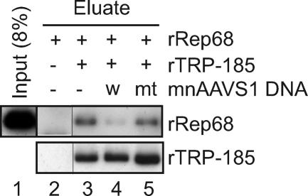
Adeno-associated virus (AAV) integrates site specifically into the AAVS1 locus on human chromosome 19. Although recruitment of the AAV nonstructural protein Rep78/68 to the Rep binding site (RBS) on AAVS1 is thought to be an essential step, the mechanism of the site-specific integration, particularly, how the site of integration is determined, remains largely unknown. Here we describe the identification and characterization of a new cellular regulator of AAV site-specific integration. TAR RNA loop binding protein 185 (TRP-185), previously reported to associate with human immunodeficiency virus type 1 TAR RNA, binds to AAVS1 DNA. Our data suggest that TRP-185 suppresses AAV integration at the AAVS1 RBS and enhances AAV integration into a region downstream of the RBS. TRP-185 bound to Rep68 directly, changing the Rep68 DNA binding property and stimulating Rep68 helicase activity. We present a model in which TRP-185 changes the specificity of the AAV integration site from the RBS to a downstream region by acting as a molecular chaperone that promotes Rep68 complex formation competent for 3'-->5' DNA helicase activity.


Figures

FIG. 1.
FIG. 1.
Identification of TRP-185 as an AAVS1-binding factor. (A) Wild-type and mutant mnAAVS1 DNA sequences and schematic representation of DNA-immobilized latex beads. (B) Input NE (lane 1), eluate from control beads (lane 2), and eluate from wild-type (lane 3) or mutant (lane 4) mnAAVS1-immobilized beads were separated on a 5 to 20% SDS-polyacrylamide gel, and proteins were visualized by silver staining. The protein bands that were differentially purified by wild-type mnAAVS1-conjugated beads are marked by dots. In the lower panel, the same samples were immunoblotted using anti-TRP-185 monoclonal antibody (NK5.18, a gift of Richard B. Gaynor). The positions of molecular mass markers are shown on the left. (C) HeLa cells were transiently transfected with pcTRP-185-Flag or pcDNA3.1(+) (mock) and then subjected to ChIP analysis using anti-Flag antibody. Input and immunoprecipitated (Ppt) DNAs were subjected to real-time PCR analysis using primers that specifically amplified the AAVS1 RBS region or a control region on chromosome 2q34. PCR products were analyzed by 2% agarose gel electrophoresis. Data represent the means ± standard errors of the mean from six independent experiments, and statistical significance, indicated by a bracket, was determined by Student's t test (P < 0.05).
FIG. 2.
FIG. 2.
Rep68 mediates AAV-AAVS1 junction formation specifically at the RBS in vitro. (A) Schematic diagrams of the AAV genome and the plasmid pBSAAVS1 containing a 1.6-kb AAVS1 sequence (left) and primer sets (right) used for in vitro junction formation assays. The positions of the primers used are indicated in the diagrams. (B) rRep68 used in this study was visualized by silver staining. The positions of molecular mass markers are shown on the right. (C) In vitro junction formation assays were performed with or without 1.5 pmol of rRep68. The recombination products and the indicated amounts of PCR standards were subjected to PCR analysis with various primer sets as shown in panel A. The PCR products were analyzed by 2% agarose gel electrophoresis and stained with ethidium bromide. The positions of size markers are shown on the right. (D) Junction formation assays were performed with the indicated amounts of rRep68 and wild-type (w) or mutant (mt) pBSAAVS1. The recombination products were examined by PCR with primer set 1 (A). The positions of size markers are shown on the right. Quantitation of the results from three independent experiments is shown below. Data represent the means ± standard errors of the mean. (E) AAV-AAVS1 junction sequences from eight clones. The RBS-flanking regions of the two substrates are shown above. In the junction sequences, overlapping sequences between AAV and AAVS1 are indicated by open boxes, and respective junction points are indicated by numbers according to the published numbering system (16). The RBS sequences are indicated by letters. nt, nucleotide.
FIG. 3.
FIG. 3.
TRP-185 inhibits Rep68-mediated AAV-AAVS1 junction formation at the RBS in vitro. (A) rTRP-185 used in this study was visualized by silver staining. The positions of molecular mass markers are shown on the right. (B) Junction formation assays were performed with the indicated amounts of rRep68, rTRP-185, heat-denatured rTRP-185 (h.d. rTRP-185), and BSA. The positions of size markers are shown on the right. Quantitation of the results from three independent experiments is shown below. Data represent the means ± standard errors of the mean. (C) Immunoblot analysis of HeLa NE depleted of TRP-185. HeLa NE was repeatedly (one to three times) passed over protein A-Sepharose to which either anti-TRP-185 or preimmune serum (ctrl) was absorbed. The supernatants were analyzed for the presence of TRP-185 and HMG-1 by immunoblotting. Quantitation of each protein level is shown below. (D) AAV-AAVS1 junction formation reactions were performed in the presence or absence of control (ctrl) or TRP-185-depleted (deltaTRP) NE (8 μg) and the indicated amounts of rRep68 and rTRP-185. The positions of size markers are shown on the right. Quantitation of the results from three independent experiments is shown below. Data represent the means ± standard errors of the mean.
FIG. 4.
FIG. 4.
TRP-185 alters AAV integration sites from the RBS to a downstream region in vivo. (A, B) HeLa cells were infected with a lentiviral expression vector encoding an shRNA that targeted TRP-185 (deltaTRP) or a control vector (ctrl). Seven days postinfection, whole cell extracts were prepared and immunoblotted with anti-TRP-185 and anti-β-tubulin antibodies (A). Alternatively, total RNA was prepared, and the mRNA level (arbitrary units) of TRP-185 was quantified by real-time RT-PCR (B). (C) In vivo integration assays were performed in control and deltaTRP HeLa cells. Following AAV or mock infection, AAV integration into the RBS, a region downstream of the RBS, and Alu repeat regions was analyzed by PCR using specific primer sets for each region, followed by Southern blot analysis. AAV-infected samples were analyzed in duplicate. The positions of DNA size markers are shown on the right. (D) Sequence analysis of AAV-AAVS1 junctions at the RBS (in deltaTRP cells) and downstream of the RBS (in control cells). In the top diagram, the positions of the PCR primers and Southern probe used are indicated, and integration events are denoted by arrows. nt, nucleotide. In the RBS junction sequences, the RBS sequences are indicated by letters, and overlapping sequences between AAV and AAVS1 are indicated by open boxes. In the downstream junction sequences, overlapping sequences between AAV and AAVS1 are indicated by letters.
FIG. 5.
FIG. 5.
TRP-185 promotes Rep68 helicase activity in an RBS-dependent manner. (A) Wild-type rRep68 and its K340H mutant were visualized by Coomassie staining. The positions of molecular mass markers are shown on the left. (B to F) Helicase assays were performed using 12.5 fmol of 32P-labeled linear AAVS1 substrate containing wild-type (B, C) or mutant (D) RBS or 10 fmol of M13 circular substrate (E, F) and the indicated amounts of rRep68 and rTRP-185. The products were then electrophoresed on a nondenaturing polyacrylamide gel. “Boil” samples were heated to 98°C for 5 min immediately before electrophoresis. Quantitation of the unwound products is shown below each panel. Schematic structures of the substrates and the unwound products are shown between or to the right of the gels.
FIG. 6.
FIG. 6.
Rep68 and TRP-185 do not bind to AAVS1 DNA simultaneously. (A, B) Gel-shift assays were performed with 10 fmol of 32P-labeled mnAAVS1 wild-type probe and the indicated amounts of rRep68 and rTRP-185. In (B), anti-His (H) and anti-Flag (F) antibodies were included in the binding reaction, as indicated (−, absent; +, present). The reaction mixtures were electrophoresed on a 4% nondenaturing polyacrylamide gel. The asterisks indicate the positions of the wells. (C) rTRP-185, rRep68, and mnAAVS1 DNA were incubated either individually or in combination and then subjected to gel filtration analysis. Fractionated samples and input materials were analyzed by immunoblotting using anti-Flag and anti-His antibodies for rRep68 and rTRP-185, respectively, or by Southern blotting using 32P-labeled mnAAVS1-specific probe. The positions of molecular mass markers are shown below. (D) The indicated amounts of rTRP-185 and rRep68 were examined, either individually or in combination, for AAVS1 binding using latex beads onto which wild-type mnAAVS1 DNA was immobilized. Eluted proteins were visualized by silver staining. The positions of molecular mass markers are shown on the left.
FIG. 7.
FIG. 7.
TRP-185 directly interacts with Rep68. rTRP-185 (1.2 pmol) was coupled to Ni-NTA beads and incubated with 3.75 pmol of rRep68 in the absence (−) or presence (+) of 2.5 pmol of mnAAVS1 wild-type (w) or mutant (mt) DNA. Eluted samples and input material were subjected to immunoblot analysis using anti-Flag and anti-His antibodies for rRep68 and rTRP-185, respectively.

Similar articles

Cited by

References

    1. Bouyac-Bertoia, M., J. D. Dvorin, R. A. Fouchier, Y. Jenkins, B. E. Meyer, L. I. Wu, M. Emerman, and M. H. Malim. 2001. HIV-1 infection requires a functional integrase NLS. Mol. Cell 7:1025-1035. - PubMed
    1. Cathomen, T., T. H. Stracker, L. B. Gilbert, and M. D. Weitzman. 2001. A genetic screen identifies a cellular regulator of adeno-associated virus. Proc. Natl. Acad. Sci. USA 98:14991-14996. - PMC - PubMed
    1. Costello, E., P. Saudan, E. Winocour, L. Pizer, and P. Beard. 1997. High mobility group chromosomal protein 1 binds to the adeno-associated virus replication protein (Rep) and promotes Rep-mediated site-specific cleavage of DNA, ATPase activity and transcriptional repression. EMBO J. 16:5943-5954. - PMC - PubMed
    1. Dignam, J. D., R. M. Lebovitz, and R. G. Roeder. 1983. Accurate transcription initiation by RNA polymerase II in a soluble extract from isolated mammalian nuclei. Nucleic Acids Res. 11:1475-1489. - PMC - PubMed
    1. Dutheil, N., M. Yoon-Robarts, P. Ward, E. Henckaerts, L. Skrabanek, K. I. Berns, F. Campagne, and R. M. Linden. 2004. Characterization of the mouse adeno-associated virus AAVS1 ortholog. J. Virol. 78:8917-8921. - PMC - PubMed

Publication types

LinkOut - more resources