. 2007 Feb;81(4):1888-98.
doi: 10.1128/JVI.02051-06. Epub 2006 Dec 6.

Murine MusD retrotransposon: structure and molecular evolution of an "intracellularized" retrovirus

Affiliations

Murine MusD retrotransposon: structure and molecular evolution of an "intracellularized" retrovirus

David Ribet et al. J Virol. 2007 Feb.

Abstract

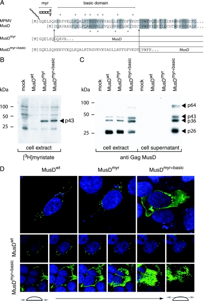
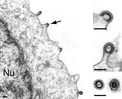
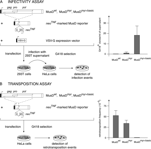
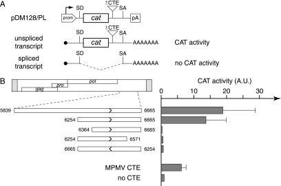
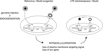
We had previously identified active autonomous copies of the MusD long terminal repeat-retrotransposon family, which have retained transpositional activity. These elements are closely related to betaretroviruses but lack an envelope (env) gene. Here we show that these elements encode strictly intracellular virus-like particles that can unambiguously be identified by electron microscopy. We demonstrate intracellular maturation of the particles, with a significant proportion of densely packed cores for wild-type MusD but not for a protease mutant. We show that the molecular origin of this unexpected intracellular localization is solely dependent on the N-terminal part of the Gag protein, which lacks a functional sequence for myristoylation and plasma membrane targeting: replacement of the N-terminal domain of the MusD matrix protein by that of its closest relative-the Mason-Pfizer monkey virus-led to targeting of the MusD Gag to the plasma membrane, with viral particles budding and being released into the cell supernatant. These particles can further be pseudotyped with a heterologous envelope protein and become infectious, thus "reconstituting" a functional retrovirus prone to proviral insertions. Consistent with its retroviral origin, a sequence with a constitutive transport element-like activity can further be identified at the MusD 3' untranslated region. A molecular scenario is proposed that accounts for the transition, during evolution, from an ancestral infectious betaretrovirus to the strictly intracellular MusD retrotransposon, involving not only the loss of the env gene but also an inability to escape the cell--via altered targeting of the Gag protein--resulting de facto in the generation of a very successful "intracellularized" insertional mutagen.


Figures

FIG. 1.
FIG. 1.
Structure of the MusD genome and associated VLPs. (A) Genomic organization of autonomous MusD elements, with the LTRs (gray) bordering the three ORFs homologous to the retroviral gag, pro, and pol genes. No ORF for an envelope protein can be identified. (B to D) Electron microscopy of the VLPs encoded by the retrotransposition competent MusD-6 copy (B and C) and a MusD-6 protease mutant (D), observed in human cells 48 h after transfection. (B) Representative low-magnification image of a transfected HeLa cell, with numerous particles organized in intracellular clusters. The nucleus (Nu), mitochondria (M), and plasma membrane (Pm) are indicated. No particle can be observed at the level of the cell membrane or in the extracellular space. (C) High-magnification view of a cluster of particles with two different morphologies: immature (solid arrow) with an electron-lucent core and mature (open arrow) with a condensed central core. (D) Same magnification as in panel C for a cluster of particles observed in cells transfected with a protease-deficient MusD-6 mutant. Only immature particles can be observed (arrow). (E) Western blot analysis of whole-cell lysates of HeLa cells transfected with the MusD-6 copy (wt, lane 1) or the protease-deficient mutant (pro, lane 2) used in panels B to D. The proteins were separated by SDS-PAGE, and Gag products were revealed using a rabbit antiserum directed against a MusD Gag recombinant protein (30). The molecular masses of the Gag precursor and protein cleavage products are indicated in kilodaltons. (F and G) Immunogold labeling of MusD particles in transfected cells using the anti-Gag MusD antiserum and a secondary antibody linked to gold beads, observed by electron microscopy; labeling is seen within the clusters (F), at the level of the MusD particles (a higher-magnification view is seen in panel G [see arrow]).
FIG. 2.
FIG. 2.
Consequences of restoring myristoylation and membrane targeting signals on MusD Gag protein addressing. (A) Sequence of the N-terminal domain of the MusD Gag protein and comparison with the corresponding domain from its closest relative, the type-D MPMV retrovirus. The consensus sequence required for myristoylation (myr, [M]GXXXS/T) and the MPMV domain rich in basic residues (basic domain, +) are indicated. Arrows indicate the limits of the replacements between the N-terminal residues of MPMV and MusD Gag proteins to generate the MusDmyr and MusDmyr+basic proviruses, respectively. (B) Restoration of MusD Gag myristoylation. Whole-cell lysates were prepared from human 293T cells transfected with the indicated MusD constructs and labeled with [3H]myristate. Myristoylated proteins were revealed by fluorography after separation by SDS-PAGE. (C) Western blot analysis of whole-cell lysates or cell supernatants from 293T cells transfected as in panel B with the indicated MusD constructs, using a rabbit antiserum directed against the MusD Gag polyprotein. The molecular masses of the Gag-specific bands are indicated in kilodaltons, as in Fig. 1E. (D) Immunofluorescence confocal analysis of human HeLa cells transfected with the indicated MusD constructs. Cells were grown on glass coverslips approximately 48 h posttransfection, fixed, and permeabilized, and MusD Gag proteins were detected by using the same rabbit anti-Gag MusD antiserum as in panel C and an Alexa Fluor 488-conjugated anti-rabbit secondary antibody. Nuclei were stained with TO-PRO-3 iodide. The two rows at the bottom show the successive confocal images of stained cells.
FIG. 3.
FIG. 3.
Plasma membrane budding and release of MusD particles upon restoration of the Gag myristoylation signal and basic domain. Electron microscopy analysis of human 293T cells transfected with the MusDmyr+basic construct. Same experimental conditions as in Fig. 1B and C. A low-magnification image is shown on the left with budding particles (arrow), and enlarged views are shown on the right, with two free particles (immature and mature) in the cell supernatant (bottom image). Bars, 0.1 μm.
FIG. 4.
FIG. 4.
Functional characterization of MusD and its variants with modified myristoylation and membrane targeting signals. (A) Infectivity assay. 293T cells were transiently transfected with an expression vector for the indicated MusD constructs and a neoTNF-marked defective MusD reporter (in which a “backward” neomycin resistance gene [with the neo ORF interrupted by a “forward” TNF intron] becomes functional only after a complete replicative cycle) (30), plus an expression vector for the VSV-G envelope protein. Supernatants from the transfected cells were collected 48 h posttransfection and used to infect naive target HeLa cells. After a 3-day growth period, infection events were detected upon G418 selection of the cells and quantitated by counting the number of G418r clones. No clone was detected in the absence of the VSV-G expression vector. (B) Transposition assay. HeLa cells were transfected with the same MusD plasmids as in panel A but without the VSV-G envelope protein expression vector; after a 6-day growth period, the cells were seeded (5 × 10⁵ cells per plate) and subjected to G418 selection to detect retrotransposition events (number of G418r clones per cells in selection [see reference 30]).
FIG. 5.
FIG. 5.
Identification of a sequence with a CTE-like activity in the MusD 3′ untranslated region. (A) Rationale of the assay and structure of the reporter pDM128/PL vector. pDM128/PL contains a cat gene flanked by donor and acceptor splice sites (SD and SA) and placed under the control of a simian virus 40 promoter and polyadenylation signal (prom, pA). Sequences to be tested for CTE activity are inserted as indicated. The presence of a CTE should promote the export of unspliced RNA from the nucleus of cells transfected with the reporter plasmid, leading to a detectable CAT activity. The absence of CTE-like activity should lead to the export of spliced RNA and a lack of CAT activity. (B) 293T cells were transiently transfected with pDM128/PL vectors containing the indicated MusD fragments, placed in the forward or reverse orientation (termini are indicated by nucleotide positions in the MusD sequence, and orientation is indicated by arrows). At 48 h posttransfection, cells were lysed and CAT activity was determined by using [14C]chloramphenicol as described in Materials and Methods. The mean ratio of CAT activity between pDM128/PL containing a MusD (or control MPMV CTE) sequence versus the empty vector (no CTE) was calculated from two to four independent experiments.
FIG. 6.
FIG. 6.
Model for the “intracellularization” of infectious retroviruses. A bona fide infectious retrovirus, e.g., the MusD progenitor, is endogenized upon infection of the germ line of a remote ancestor and Mendelian transmission to the following generations, thus resulting in a so-called ERV. This ERV may retain the characteristic features of retroviruses, i.e., produce infectious extracellular particles with a functional envelope protein, and be prone to horizontal transmission. Intracellularization is expected to correspond to an additional adaptation of the progenitor retrovirus, in which the produced VLPs are no longer able to exit or reenter the cell. For MusD, this has been achieved via the loss of the plasma membrane targeting function (degeneration of the myristoylation signal and basic domain at the N-terminal end of the matrix Gag protein) and the loss of the env gene. The resulting strictly intracellular life cycle resembles that of primitive Ty-like LTR-retrotransposons (although both classes of elements should be evolutionarily distinct) and is associated—at least in the case of the MusD element—with high-efficiency retrotransposition.

Similar articles

Cited by

References

    1. Baillie, G. J., L. N. van de Lagemaat, C. Baust, and D. L. Mager. 2004. Multiple groups of endogenous betaretroviruses in mice, rats, and other mammals. J. Virol. 78:5784-5798. - PMC - PubMed
    1. Bannert, N., and R. Kurth. 2004. Retroelements and the human genome: new perspectives on an old relation. Proc. Natl. Acad. Sci. USA 13:14572-14579. - PMC - PubMed
    1. Barbulescu, M., G. Turner, M. I. Seaman, A. S. Deinard, K. K. Kidd, and J. Lenz. 1999. Many human endogenous retrovirus K (HERV-K) proviruses are unique to humans. Curr. Biol. 9:861-868. - PubMed
    1. Baust, C., G. J. Baillie, and D. L. Mager. 2002. Insertional polymorphisms of ETn retrotransposons include a disruption of the wiz gene in C57BL/6 mice. Mamm. Genome 13:423-428. - PubMed
    1. Beliakova-Bethell, N., C. Beckham, T. H. Giddings, Jr., M. Winey, R. Parker, and S. Sandmeyer. 2006. Virus-like particles of the Ty3 retrotransposon assemble in association with P-body components. RNA 12:94-101. - PMC - PubMed

Publication types

MeSH terms

LinkOut - more resources