. 2006 Dec 19;103(51):19326-31.
doi: 10.1073/pnas.0609436103. Epub 2006 Dec 6.

Crystal structure of apo-calmodulin bound to the first two IQ motifs of myosin V reveals essential recognition features

Affiliations

Crystal structure of apo-calmodulin bound to the first two IQ motifs of myosin V reveals essential recognition features

Anne Houdusse et al. Proc Natl Acad Sci U S A. .

Abstract

A 2.5-A resolution structure of calcium-free calmodulin (CaM) bound to the first two IQ motifs of the murine myosin V heavy chain reveals an unusual CaM conformation. The C-terminal lobe of each CaM adopts a semi-open conformation that grips the first part of the IQ motif (IQxxxR), whereas the N-terminal lobe adopts a closed conformation that interacts more weakly with the second part of the motif (GxxxR). Variable residues in the IQ motif play a critical role in determining the precise structure of the bound CaM, such that even the consensus residues of different motifs show unique interactions with CaM. This complex serves as a model for the lever arm region of many classes of unconventional myosins, as well as other IQ motif-containing proteins such as neuromodulin and IQGAPs.


Conflict of interest statement

The authors declare no conflict of interest.

Figures
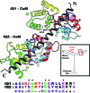
Fig. 1.
Fig. 1.
Structure of the myosin V 2IQ complex. Two CaMs bound in tandem to the two IQ motifs (gray helix) derived from the sequence adjacent to the motor domain of murine myosin V are shown. Consensus sequence residues (*) of the IQ motif are shown in ball and stick. The helices of CaM, designated A–H, are colored in pairs (AB in green, CD in yellow, EF in red, GH in cyan). The orientation of the IQ motifs and CaM are antiparallel. The interlobe linker 2 (purple) joins the N- and C-terminal lobes. Linker 1 (pink, between the B and C helices) and linker 3 (blue, between the F and G helices) interact with consensus residues of the IQ motif. (Inset) A cartoon of the myosin V molecule and the region that was crystallized (red box).
Fig. 2.
Fig. 2.
Conserved features of the CaM / IQ motif recognition. CaM bound to each IQ motif adopts a semi-open C-lobe, and a closed N-lobe. The ribbon diagrams represent CaM (color-coded as in Fig. 1) bound to the first IQ motif in two orthogonal views about the y axis. Major interactions with the semi-open C-terminal lobe are: consensus residues Gln-774 and Arg-778 (green) form five hydrogen bonds with main chain atoms in linker 3 (blue), whereas apolar side chains (Ile-773, yellow; Ile-777, purple; Trp-780, black and pink) interact within the hydrophobic C-lobe. Consensus residues Gly-779 (orange ball) and Arg-783 (cyan), as well as Tyr-786 (yellow) interact with the surface of the N-lobe composed of linker 1 (pink) and helix A. Hydrogen bonds between Glu-114 in linker 3 of the C-lobe, and the main-chain nitrogen of Glu-45 and Ala-46 in the N-lobe, provide a sensing mechanism between the two halves of CaM.
Fig. 3.
Fig. 3.
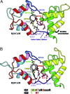
Variable residues in the IQ motif affect interactions with the interlobe linker and influence the N-lobe orientation of CaM. Ribbon diagrams of CaM bound to the first (A) and the second (B) IQ motifs display the major differences between the two complexes: a 20° difference in the orientation of the N lobe (relative to the heavy chain helix), and variability in the conformation of the interlobe linker. The last turn of helix D of CaM bound to the second IQ motif is also unfolded. Differences in the sequence of the IQ motif residues (Arg-772/Thr-795, Lys-775/Arg-798, Thr-776/Tyr-799) that interact with the interlobe linker (Asp-80, Glu-84) of CaM cause the N lobe to be further away from the IQ helix in IQ2 compared with IQ1. A key difference for the linker conformation is the substitution of Thr-776 in IQ1 for the bulkier Tyr-799 in IQ2 that interacts specifically with the end of the interlobe linker. The side-chain of Arg-772 in motif 1 forms a hydrogen bond with the sidechain of Asp-80 in the interlobe linker, whereas the smaller Thr-795 in motif 2 cannot do this. The variable Lys-775 in motif 1 forms a hydrogen-bond only with Asp-80, whereas the comparable Arg-798 in IQ2 forms hydrogen-bond with Asp-80 in the interlobe linker as well as Glu-84 in helix E.
Fig. 4.
Fig. 4.
Binding of the N-lobe of CaM to the second half of the two IQ motifs. The residues involved in N-lobe binding correspond to the second half of the IQ motif, i.e., GWLDRKRYLCMQ for motif 1 (residues 779–790) and GYQARCYAKFLR for motif 2 (residues 802–813). The N-lobes are oriented similarly to compare the interactions made with the IQ motif helix. The variable residues affect the interlobe linker conformation (see Fig. 3), and thus the relative position of the C and N lobes differ in the two complexes. The different orientations are illustrated by a red-dashed line that represents the axis of the E helix of the C lobe as found in IQ1. The gray-dashed line, which represents the axis of the IQ helix as found in IQ2, shows that the IQ helix is further away from the N lobe in motif 2 than in motif 1. Tyr-786 in motif 1 provides additional interactions with helix A that are not found in the second IQ motif, where Tyr is replaced by the smaller Ala. Gln-790 forms a hydrogen bond with Glu-14 on helix A, whereas this interaction does not occur in motif 2 because the side chain of Arg-813 is oriented such that it is ≈7 Å from Glu-14. Even the conserved consensus residue Arg-783 in motif 1 and Arg-806 in motif 2 differ in how they interact with the IQ motif peptide.
Fig. 5.
Fig. 5.
Conformational change in an EF-hand lobe when Ca2+ binds. Comparison of the C-lobe conformation in the semi-open state (Upper) and open state (Lower; ref. 15), after superimposition of the E helix. Note the difference in the orientation of the four helices (blue) as well as the difference in position of the target peptide (gray). The conformation of loop 3 (between helices E and F) is similar to that found when Ca2+ is bound, and most residues involved in Ca2+ chelation are in position to participate in binding. The major exception is Glu-104 (yellow) in helix F, which is positioned too far from the rest of loop 3 to coordinate Ca2+ in the semi-open lobe. Its recruitment in the coordination sphere requires a major change in the relative orientation of helices E and F. By contrast, the conformation of loop 4 between helices G and H (cyan, Right) in the semi-open lobe, greatly differs from that required for Ca2+ binding.

References

    1. Trybus KM. Nat Cell Biol. 2005;7:854–856. - PubMed
    1. Sellers JR, Veigel C. Curr Opin Cell Biol. 2006;18:68–73. - PubMed
    1. Wang F, Chen L, Arcucci O, Harvey EV, Bowers B, Xu Y, Hammer JA, III, Sellers JR. J Biol Chem. 2000;275:4329–4335. - PubMed
    1. Krementsov DN, Krementsova EB, Trybus KM. J Cell Biol. 2004;164:877–886. - PMC - PubMed
    1. Wang F, Thirumurugan K, Stafford WF, Hammer JA, III, Knight PJ, Sellers JR. J Biol Chem. 2004;279:2333–2336. - PubMed

Publication types

Associated data

LinkOut - more resources