Comparative Study
. 2006 Dec 19;103(51):19424-9.
doi: 10.1073/pnas.0609184103. Epub 2006 Dec 6.

Zfp423 controls proliferation and differentiation of neural precursors in cerebellar vermis formation

Affiliations
Comparative Study

Zfp423 controls proliferation and differentiation of neural precursors in cerebellar vermis formation

Wendy A Alcaraz et al. Proc Natl Acad Sci U S A. .

Abstract

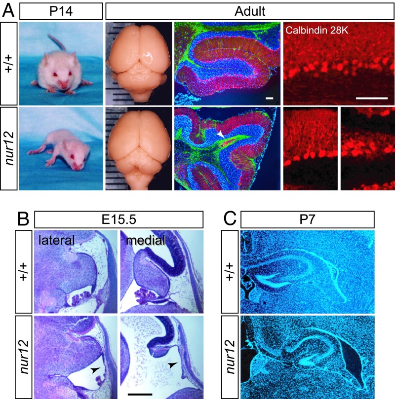
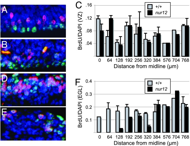
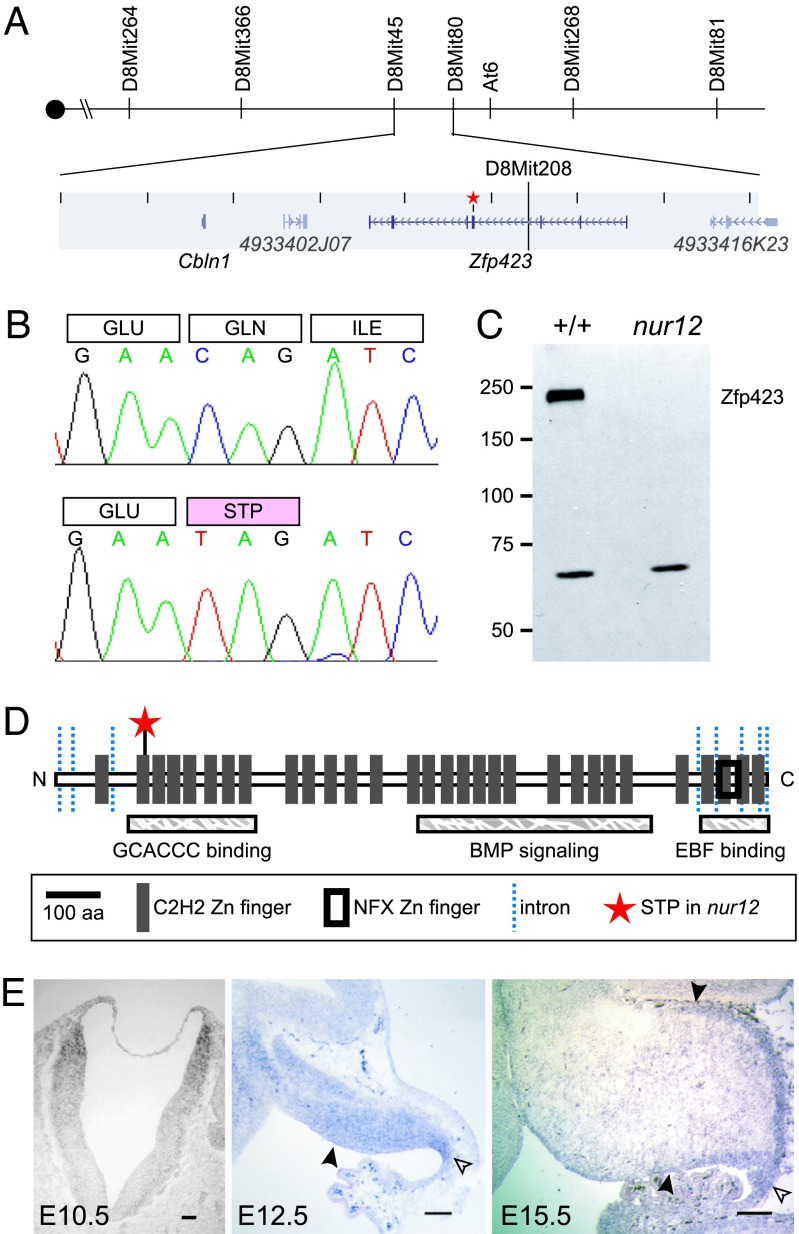
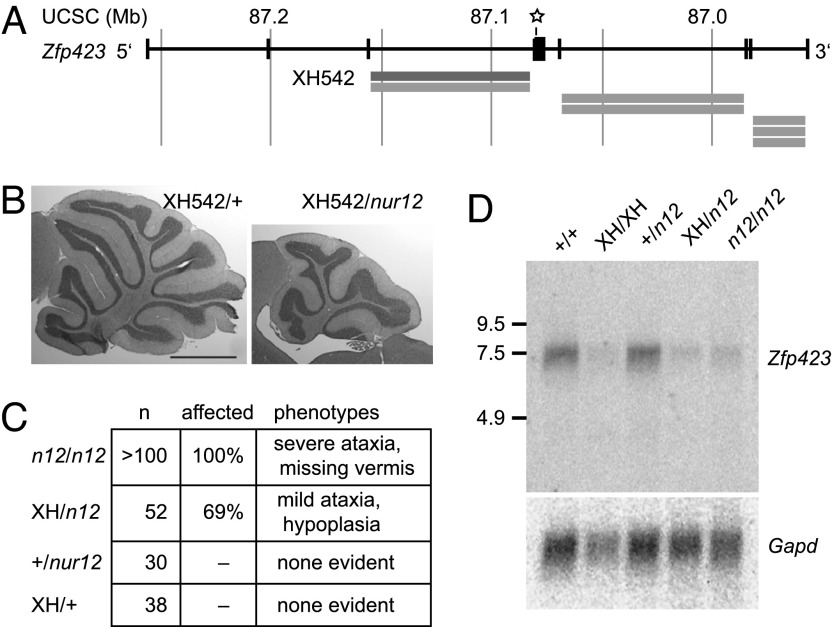
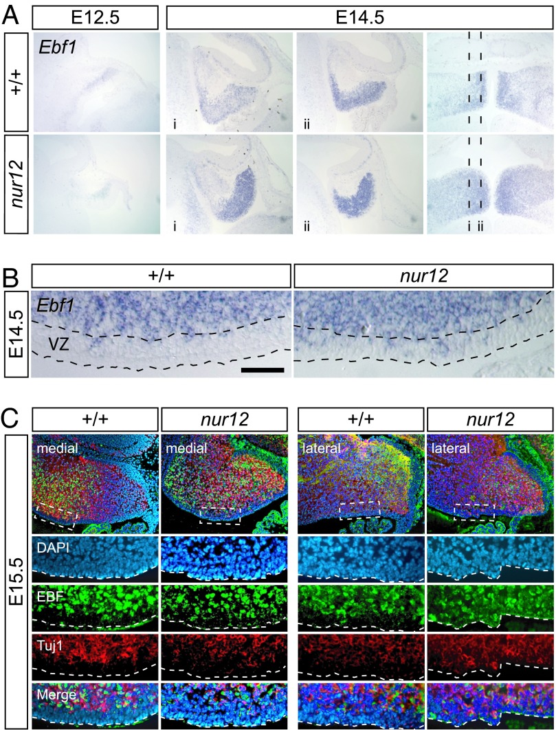
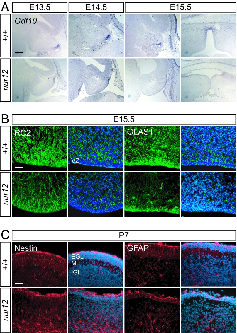
Neural stem cells and progenitors in the developing brain must choose between proliferation with renewal and differentiation. Defects in navigating this choice can result in malformations or cancers, but the genetic mechanisms that shape this choice are not fully understood. We show by positional cloning that the 30-zinc finger transcription factor Zfp423 (OAZ) is required for patterning the development of neuronal and glial precursors in the developing brain, particularly in midline structures. Mutation of Zfp423 results in loss of the corpus callosum, reduction of hippocampus, and a malformation of the cerebellum reminiscent of human Dandy-Walker patients. Within the cerebellum, Zfp423 is expressed in both ventricular and external germinal zones. Loss of Zfp423 results in diminished proliferation by granule cell precursors in the external germinal layer, especially near the midline, and abnormal differentiation and migration of ventricular zone-derived neurons and Bergmann glia.


Conflict of interest statement

The authors declare no conflict of interest.

Figures

Fig. 1.
Fig. 1.
Ataxia and malformation in nur12 mutant mice. (A) P14 littermates photographed midstride illustrate severe ataxia and reduced weight gain by nur12 mutants. Surface view of littermate brains shows selective loss of the cerebellar vermis. (Grid lines: 1 mm.) Fluorescence micrographs with Calbindin (red), GFAP (green), and DAPI (blue) illustrate diminished foliation and heterotopic clusters of Purkinje cells (arrow). Higher-magnification image shows dendritic arborization in both aligned (Left) and heterotopic (Right) cells in nur12. (Scale bars: 100 μm.) (B) Sections through E15.5 embryos show midline-biased hypoplasia of cerebellum and choroid plexus. Note the anterior rotation of the cerebellum and distortion of the area membranacea superior (ams, arrowheads). (Scale bar: 500 μm.) (C) Dysgenesis of hippocampal formation and enlargement of the lateral ventricles of nur12 mutant compared with littermate control. Nuclear DAPI stain reveals pattern of cell bodies.
Fig. 2.
Fig. 2.
Reduced proliferation rate is biased toward the midline in EGL. Immunofluorescence and summary graphs illustrate proliferation rates along the mediolateral axis of ventricular zone (AC) and external germinal zone (DF) in nur12 and control littermates. BrdU incorporation (red) and Ki67 protein expression (green) are seen among DAPI-stained nuclei (blue) in ventricular zone [wild-type (A); nur12 (B)] and external germinal layer [wild-type (D); nur12 (E)]. Representative fields 192 μm from the midline at E14.5 are shown. Graphs show the ratio of BrdU-positive nuclei to DAPI-stained nuclei in ventricular zone (C) and external germinal layer (F) as a function of distance from the midline. Data are pooled counts from matched pairs of nur12/nur12 and +/+ littermates. Standard errors of the mean are indicated. No EGL was present in at least one nur12 animal for each point from 0 to 192 μm. Single pairs were counted at 576 and 704 μm.
Fig. 3.
Fig. 3.
nur12 is a null allele of Zfp423. (A) Recombination map for nur12. D8Mit45 is excluded by one and D8Mit80 by three recombination events. The nonrecombining interval contains Cbln1, Zfp423, and two ESTs. The star marks the site of the mutation. (B) Sequencing RT-PCR products from mutant and control identifies a nonsense mutation in Zfp423, at codon 166 of 1,292 with respect to RefSeq protein NP_201584. (C) Monoclonal antibody detects Zfp423 in neonatal brain extracts from +/+ but not nur12. A cross-reacting band of unknown origin is seen in both genotypes. (D) Zfp423 encodes 30 predicted C2H2 zinc-finger domains. Domains required for consensus site binding, BMP signaling, and EBF binding (13) are labeled. The star shows the nur12 stop codon; lines indicate exon junctions. (E) In situ hybridization shows Zfp423RNA expression in dorsal neural tube of the presumptive hindbrain by E10.5. At E12.5, Zfp423 is detected in the ventricular zone (filled arrowhead) of the cerebellar anlage and in the rhombic lip (open arrowhead). At E15.5, Zfp423 is detected in the remaining ventricular zone, rhombic lip, and EGL (upper arrowhead).
Fig. 4.
Fig. 4.
A hypomorphic Zfp423 gene trap allele does not complement nur12. (A) Schematic view of Zfp423 locus showing BayGenomics gene-trap cell lines. Orientation is reversed with respect to the chromosome to show Zfp423 5′ to 3′. Reported gene-trap integration sites (localized to introns by 5′ RACE) are indicated by gray bars. The star shows the location of stop codon in nur12. (B) Cross-sectional area of the vermis of XH542/nur12 is approximately half that of XH542/+ or +/+ controls. (Scale bar: 1 mm.) (C) Distribution of cerebellar phenotypes is indicated for each Zfp423 genotype. Ataxia and vermis agenesis are fully penetrant in nur12, although involvement of hemispheres is variable. Mild ataxia and cerebellar hypoplasia are seen in XH542/nur12 but not in either single heterozygote from the same cross. (D) Gene-trap mice have reduced expression of Zfp423. Northern blot of 7 μg of total RNA for animals of the indicated genotypes hybridized with Zfp423 cDNA fragment 3′ to the insertion site. Size markers are indicated in kilobases.
Fig. 5.
Fig. 5.
Differentiation of ventricular zone neurons in nur12. (A) In situ hybridization shows Ebf1 expression in postmitotic neurons (presumptive Purkinje cells) in the cerebellum by E12.5. At E14.5, the mediolateral pattern of Ebf1 is both increased and more diffuse in nur12 than in control littermate. Approximate locations of sagittal sections are indicated on the horizontal section at right. (B) Ebf1-expressing cells in the ventricular zone (VZ) are approximately twice as frequent in nur12 than in littermate control; sagittal plane, posterior to the right. (Scale bar: 50 μm.) (C) Immunofluorescence shows EBF, Tuj1 double-positive cells in the most superficial layer of the nur12 ventricular zone at E15.5, indicating neuronal identity of these cells, in both medial and lateral sections. The proportion of EBF+, Tuj1+ cells in VZ of nur12 is approximately twice that of wild type in both medial and lateral sections.
Fig. 6.
Fig. 6.
Defects in Bergmann glial and neuroepithelial progenitor cells. (A) Gdf10, a marker for presumptive Bergmann glia, appears in ventricular zones of both nur12 and littermate controls by E13.5. At E14.5, expression is reduced in the hemispheres of the mutant. At E15.5, migrating Gdf10-positive cells are seen in the cerebellar cortex of the control but not the mutant in either sagittal (Left) or horizontal (Right) orientations. (Scale bar: 200 μm.) (B) Immunofluorescence shows fewer glial cell bodies and radial fibers in nur12. Both immature progenitors in the ventricular zone (VZ) and migrating glia express RC2 and GLAST. By E15.5, fewer marker-positive cells are seen in either VZ or presumptive cortex of nur12 compared with littermate control, although some radial fibers are evident. (Scale bar: 100 μm.) (C) By P7, nestin- and GFAP-positive cells remain poorly organized in nur12, although end feet are clearly visible at the pial surface above the EGL. Merged images show nuclei stained by DAPI. VZ, ventricular zone; EGL, external granule layer; ML, molecular layer; IGL, internal granule layer. (Scale bar: 100 μm.)

References

    1. ten Donkelaar HJ, Lammens M, Wesseling P, Thijssen HO, Renier WO. J Neurol. 2003;250:1025–1036. - PubMed
    1. Grinberg I, Northrup H, Ardinger H, Prasad C, Dobyns WB, Millen KJ. Nat Genet. 2004;36:1053–1055. - PubMed
    1. Kile BT, Hentges KE, Clark AT, Nakamura H, Salinger AP, Liu B, Box N, Stockton DW, Johnson RL, Behringer RR, et al. Nature. 2003;425:81–86. - PubMed
    1. Tsai RY, Reed RR. Mol Cell Biol. 1998;18:6447–6456. - PMC - PubMed
    1. Tsai RY, Reed RR. J Neurosci. 1997;17:4159–4169. - PMC - PubMed

Publication types

MeSH terms