. 2007 Feb;175(2):567-84.
doi: 10.1534/genetics.106.065219. Epub 2006 Dec 6.

Onset of the DNA replication checkpoint in the early Drosophila embryo

Affiliations

Onset of the DNA replication checkpoint in the early Drosophila embryo

Justin Crest et al. Genetics. 2007 Feb.

Abstract

The Drosophila embryo is a promising model for isolating gene products that coordinate S phase and mitosis. We have reported before that increasing maternal Cyclin B dosage to up to six copies (six cycB) increases Cdk1-Cyclin B (CycB) levels and activity in the embryo, delays nuclear migration at cycle 10, and produces abnormal nuclei at cycle 14. Here we show that the level of CycB in the embryo inversely correlates with the ability to lengthen interphase as the embryo transits from preblastoderm to blastoderm stages and defines the onset of a checkpoint that regulates mitosis when DNA replication is blocked with aphidicolin. A screen for modifiers of the six cycB phenotypes identified 10 new suppressor deficiencies. In addition, heterozygote dRPA2 (a DNA replication gene) mutants suppressed only the abnormal nuclear phenotype at cycle 14. Reduction of dRPA2 also restored interphase duration and checkpoint efficacy to control levels. We propose that lowered dRPA2 levels activate Grp/Chk1 to counteract excess Cdk1-CycB activity and restore interphase duration and the ability to block mitosis in response to aphidicolin. Our results suggest an antagonistic interaction between DNA replication checkpoint activation and Cdk1-CycB activity during the transition from preblastoderm to blastoderm cycles.


Figures
F<sc>igure</sc> 1.—
Figure 1.—
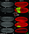
Cycle 10 (A–E) and cycle 14 (A′–E′) phenotypes. Embryos were fixed and stained with anti-histone H1 at cycle 10 (A–E). Embryos were also fixed at cycle 14 and stained with anti-histone H1 to label nuclei (red) and antiphosphorylated histone H3 (green), which labels nuclei undergoing mitosis (A′–E′). two cycB (wild-type) embryos at cycle 10 (A) and cycle 14 (A′) are shown. Six CycB embryos at cycle 10 (B) and cycle 14 (B′) are shown. Arrows in B and E indicate nuclei, which have not reached the cortex at cycle 10. Note the patch of anti-phospho-histone H3, indicating nuclei in M-phase (B′ and D′); inset in B′′ shows chromosomal bridges. An example of a class I suppressing Df at cycle 10 (C) and cycle 14 (C′) is shown. (D and D′) A case of a class II suppressing gene (Arp87C). (E and E′) An example of a class III suppressing Df. Pole cells (p) are in M-phase at cycle 14 in both six cycB (B′) and class II suppressing embryos (D′).
F<sc>igure</sc> 2.—
Figure 2.—
Cytogenetic map of polytene salivary gland chromosomes with deficiencies indicating modification of the six cyclin B phenotype. The map includes data from our previous screen (open bars, Ji et al. 2002) and from the extended screen reported here (solid bars). The Drosophila euchromatic genome consists of four chromosomes, of which three are represented here with each of the five chromosomal arms (X, 2L, 2R, 3L, and 3R) subdivided into 20 salivary gland units (100 units total). Relative lengths of deficiencies tested for modification are shown. Black bars indicate deficiencies that neither enhanced nor suppressed the six cycB phenotype. Red bars are deficiencies that enhanced and blue bars are those that suppressed the six cycB phenotype. Specific genes that were identified as modifiers are colored similarly; dRPA2 (CG9273) was a suppressor identified in this screen. x indicates a previous falsely identified enhancer that after retesting with a molecularly defined Df in region 61 failed to enhance the six cycB phenotype. dRPA1 Df (84F6) did not enhance or suppress the six cycB phenotype. For specific breakpoints of the deficiencies tested here see the appendix.
F<sc>igure</sc> 3.—
Figure 3.—
Images from a single embryo used to define interphase duration (A and A′), prophase–metaphase duration (B and B′), and anaphase–telophase duration (C and C′). Interphase (i) begins 10 sec (A) after the latest telophase configuration (t in C′). Note the pair of teardrop-shaped nuclei (t), 10 sec before earliest interphase (A). Interphase ends 10 sec before earliest prophase (p in B; note loss of round nuclear morphology). Prophase–metaphase duration (B–B′) begins when first condensation is observed (p in B) and ends 10 sec before anaphase (a in B′). Metaphase (m) occurs within this duration. Anaphase–telophase duration (C–C′) begins when first movement of chromatids is observed (a in C, note the weak signal from sister chromatids below indicated with a dotted line) and ends in late telophase (t in C′).
F<sc>igure</sc> 4.—
Figure 4.—
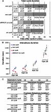
Cell-cycle (A) and cell-cycle phase (B and C) durations during the syncytial blastoderm divisions with varying doses of Cyclin B and the suppression of six cycB by dRPA2. Cell-cycle and phase durations were defined using a Hist2AvD-EGFP tag and two-photon live imaging. For each genotype, five to seven embryos were recorded and analyzed. In all four cycles, one cycB embryos had longer cycles (A) and interphases (B), but shorter prophase–metaphase durations (C) than two cycB embryos, while six cycB embryos had the shortest cycles (A) and interphase durations (B), but longest prophase–metaphase durations (C). Anaphase–telophase durations showed no differences among the genotypes (C). Reducing dRPA2 gene copy number extended interphase more in later blastoderm cycles (B). Looking at total cell-cycle time (A) no differences were observed compared to six cycB embryos. Arrows in B indicate no statistical differences from six cycB (red) or two cycB (black). [P ≤ 0.002, in all cases, on the basis of the isotonic regression test (Gaines and Rice 1990).] *Cell-cycle phases presented in seconds. Significant difference from two cycB (based on Tukey–Kramer means comparison test, P < 0.001; Sokal and Rohlf 1981).
F<sc>igure</sc> 5.—
Figure 5.—
Blockage of DNA replication tests for checkpoint effect in embryos with different amounts of CycB and dRPA2. (A) Control and experimental embryos (x-axis) were injected at 60, 90, 120, or 135 min AED (z-axis). Control embryos were injected with 1% DMSO, experimental embryos with 0.1% aph. solution in 1% DMSO, and both were fixed 30 min later. Embryos were scored as normal (y-axis) if they contained no nuclei with bridges or other division defects; abnormal embryos had at least five nuclei with defects described in the text. *Significant differences in one cycB and six cycB when compared to aph.-injected two cycB embryos. Significant differences in dRPA21 embryos compared to their respective cycB controls (P ≤ 0.001). (B) Recordings of single embryos using His2AvD-EGFP and TPLSM microscopy. One (blue), two (black), and six (red) cycB embryos were injected during late prophase of cycles 9 or 11. Recording started (0 min) in early interphase (inter) of cycles 10 or 12, respectively, and until the last moment of interphase. To illustrate checkpoint activity images from recordings are presented here at 5-min intervals within each interphase and ending with the last interphase (inter). Representative cases from each genotype were chosen and their actual interphase duration times are presented here.
F<sc>igure</sc> 6.—
Figure 6.—
Histone H3 dephosphorylation begins earlier in anaphase of a six cycB embryo than in that of one, two cycB, and dRPA2/six cycB embryos. Late anaphase of one cycB (A, A′), two cycB (B, B′), and dRPA2/six cycB (D, D′) and early anaphase of six cycB (C, C′) embryos fixed at cycle 10 and stained with an antibody to histone H1 (red) and the phosphorylated form of histone H3 (green) are shown. Note the earlier loss of PH3 in six cycB nuclei (C, C′) in 6 of 10 cases observed as compared to the later anaphase of one cycB in 4 of 4 cases, of two cycB (B, B′) in 5 of 5 cases, and of dRPA2/six cycB (D, D′) in 4 of 4 cases. This indicates that six cycB embryos have delayed Grp activity when compared with two cycB (Su et al. 1999).

References

    1. Blankenship, J. T., and E. Wieschaus, 2001. Two new roles for the Drosophila AP patterning system in early morphogenesis. Development 128: 5129–5138. - PubMed
    1. Blumenthal, A. B., H. J. Kriegstein and D. S. Hogness, 1974. The units of DNA replication in Drosophila melanogaster chromosomes. Cold Spring Harbor Symp. Quant. Biol. 38: 205–223. - PubMed
    1. Brodsky, M. H., J. J. Sekelsky, G. Tsang, R. S. Hawley and G. M. Rubin, 2000. mus304 encodes a novel DNA damage checkpoint protein required during Drosophila development. Genes Dev. 14: 666–678. - PMC - PubMed
    1. Clarkson, M., and R. Saint, 1999. A His2AvDGFP fusion gene complements a lethal His2AvD mutant allele and provides an in vivo marker for Drosophila chromosome behavior. DNA Cell Biol. 18: 457–462. - PubMed
    1. Costanzo, V., D. Shechter, P. J. Lupardus, K. A. Cimprich, M. Gottesman et al., 2003. An ATR- and Cdc7-dependent DNA damage checkpoint that inhibits initiation of DNA replication. Mol. Cell 11: 203–213. - PubMed

Publication types

MeSH terms

LinkOut - more resources