Comparative Study
. 2006 Dec 6;26(49):12735-47.
doi: 10.1523/JNEUROSCI.0575-06.2006.

Neuronal pentraxin 1 contributes to the neuronal damage evoked by amyloid-beta and is overexpressed in dystrophic neurites in Alzheimer's brain

Affiliations
Comparative Study

Neuronal pentraxin 1 contributes to the neuronal damage evoked by amyloid-beta and is overexpressed in dystrophic neurites in Alzheimer's brain

Maria A Abad et al. J Neurosci. .

Abstract

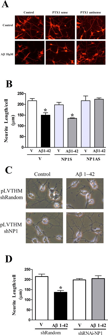
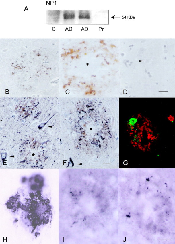
Accumulation of amyloid-beta (Abeta) is thought to play a central role in the progressive loss of synapses, the neurite damage, and the neuronal death that are characteristic in brains affected by Alzheimer's disease. However, the mechanisms through which Abeta produces such neurotoxicity remain unclear. Because Abeta depresses synaptic activity, we investigated whether the neurotoxicity of Abeta depends on the expression of NP1, a protein involved in excitatory synapse remodeling that has recently been shown to mediate neuronal death induced by reduction in neuronal activity in mature neurons. We found that treatment of cortical neurons in culture with Abeta produces a marked increase in NP1 protein that precedes apoptotic neurotoxicity. Silencing NP1 gene expression by RNA interference (short hairpin RNA for RNA interference) prevents the loss of synapses, the reduction in neurite outgrowth, and the apoptosis evoked by Abeta. Transgene overexpression of NP1 reproduced these neurotoxic effects of Abeta. Moreover, we found that NP1 was increased in dystrophic neurites of brains from patients with sporadic late-onset Alzheimer's disease. Dual immunohistochemistry for NP1 and tau showed that NP1 colocalizes with tau deposits in dystrophic neurites. Furthermore, NP1 colocalized with SNAP-25 (synaptosomal-associated protein of 25 kDa) in the majority of dystrophic neurites surrounding amyloid deposits. NP1 was also increased in cell processes surrounding amyloid plaques in the cerebral cortex and hippocampus of APP/PS1 (mutant amyloid precursor protein/presenilin 1) transgenic mice. These findings show that NP1 is a key factor for the synapse loss, the neurite damage, and the apoptotic neuronal death evoked by Abeta and indicate that Abeta contributes to the pathology of Alzheimer's disease by regulating NP1 expression.


Figures

Figure 1.
Figure 1.
Oligomeric Aβ1–42 induces apoptotic neuronal death. Cortical neuronal cultures (4–6 DIV) were treated with different concentrations of oligomeric Aβ1–42. A, Soluble Aβ1–42 increases the percentage of condensed nuclei in a concentration-dependent manner. Condensed nuclei were counted after Hoechst 33258 staining 48 h after treatment. V, Vehicle. B, Time course of neurotoxicity induced by soluble Aβ1–42 (10 μm). In these experiments, neuronal death was measured by counting the number of PI stained over total number of cells in digital images of fluorescence and phase-contrast photomicrographs simultaneously obtained in an inverted fluorescence microscope. C, Control. C, Representative photomicrographs of cortical neuronal cultures treated with either vehicle or soluble Aβ1–42 (10 μm) for 48 h and stained with Hoechst 33258 (top) or after immunocytochemistry for cleaved caspase 3 (bottom). Arrowheads indicate condensed nuclei. The data shown are from three independent experiments and are expressed as a percentage of the values in control cultures. *p < 0.05, significantly different from the corresponding control (one-way ANOVA with Bonferroni post hoc comparisons).
Figure 2.
Figure 2.
Oligomeric Aβ1–42 increases NP1 expression in cortical neuronal cultures. A, Representative Western blot showing the effect of a maximal concentration of oligomeric Aβ1–42 (20 μm) on NP1 and actin levels over time. B, Quantitative analysis showing a concentration-dependent effect of oligomeric Aβ1–42 (10 and 20 μm) on NP1 protein levels. Western blots were incubated with mouse anti-NP1 antibody (1:1000). Densitometric values of the bands representing NP1 immunoreactivity were normalized with the values of the corresponding actin bands. The ratio of NP1 over actin was expressed as a percentage of the control values. Values are the mean ± SEM of at least three independent experiments. *p < 0.05, significantly different from control values (one-way ANOVA with Bonferroni post hoc comparisons). C, Control.
Figure 3.
Figure 3.
Silencing NP1 expression rescues cortical neurons from apoptosis evoked by Aβ. Expression of NP1 was silenced with antisense ODNs or with lentiviral-mediated RNAi. A, B, Cortical neuronal cultures were transfected with either antisense (NP1AS) or the corresponding sense (NP1S) ODN (0.8 μg) at 4 DIV, as described in Materials and Methods. Approximately 4 h later, neurons were treated with vehicle or oligomeric Aβ1–42 (10 μm). A, Apoptotic nuclei were measured with Hoechst 33258 staining 48 h after treatment with Aβ. B, The percentage of neurons with cleaved caspase 3 was measured by immunocytochemistry using a rabbit anti-human cleaved caspase-3 polyclonal antiserum 48 h after treatment with 10 μm Aβ1–42. C, Cortical neuronal cultures were transduced with a control lentivirus vector expressing a random sequence (pLVTHM-shRandom) or with a lentivirus vector expressing a short hairpin sequence that produces a small interfering RNA directed against NP1 mRNA (pLVTHM-shRNAi-NP1). Ten to 20 μl of viral lentivirus particle stock were added at the same time of plating. The cells were treated with the vehicle or soluble Aβ1–42 (10 μm) at 4 DIV, and apoptotic nuclei were measured with Hoechst 33258 staining 48 h after treatment with Aβ. D, Representative Western blot showing a reduction in NP1 protein levels after exposure to lentivirus-mediated RNAi. Proteins were extracted 18 h after treatment with Aβ1–42, separated on 10% SDS-PAGE, and transferred to PVDF membranes. Western blots were incubated with mouse anti-NP1 antibody (1:1000). *p < 0.05, significantly different from control values (one-way ANOVA with Bonferroni post hoc comparisons). Values are mean ± SE of three independent experiments. V, Vehicle; C, control.
Figure 4.
Figure 4.
Silencing NP1 expression prevents the reduction in neurite outgrowth evoked by Aβ. Expression of NP1 was silenced with antisense ODNs or with lentiviral-mediated RNAi. A, B, Cortical neurons, plated at low density in Permanox coverslides, were transfected with either antisense (NP1AS) or the corresponding sense (NP1S) ODN (0.8 μg) at 1 DIV. Approximately 4 h later, neurons were treated with vehicle or soluble Aβ1–42 (10 μm) and fixed with 4% paraformaldehyde 48 h after treatment with Aβ. A, Representative immunofluorescence images of the tubulin network of cortical neuronal cultures assayed with anti-α-tyrosinated-tubulin monoclonal antibody. B, Total neurite length was estimated in photomicrographs of the tubulin network using a stereological procedure. C, Phase-contrast representative photomicrographs of cortical neurons transduced with a control lentivirus (pLVTHM-shRandom) or a lentivirus vector producing an NP1-specific small interfering RNA (pLVTHM-shRNAi-NP1). Lentivirus particles were added at the same time of plating, and treatment with vehicle or soluble Aβ1–42 (10 μm) was performed at 1 DIV. D, Total neurite length was estimated from phase-contrast photomicrographs obtained from living cells 24 h after treatment with Aβ using a stereological procedure (Ronn et al., 2000). Values are mean ± SE of three independent experiments. *p < 0.05, significantly different from control values (one-way ANOVA with Bonferroni post hoc comparisons). V, Vehicle.
Figure 5.
Figure 5.
Silencing NP1 expression prevents synapse loss evoked by Aβ. Expression of NP1 was silenced with lentiviral-mediated RNAi. Cortical neuronal cultures were transduced with the control lentiviral vector pLVTHM-shRandom or with the lentiviral vector for RNAi against NP1, pLVTHM-shRNAi-NP1. Five microliters of viral lentivirus particle stock were added at the same time of plating. The cells were treated with vehicle or oligomeric Aβ1–42 (20 μm) at 5 DIV for 48 h. A, Representative Western blot showing a reduction in synaptophysin protein levels after exposure to Aβ. Proteins were extracted 48 h after treatment with Aβ1–42. Western blots were incubated with mouse anti-synaptophysin antibody (SY38; 1:1000). Actin was used as control for protein loading. B, Quantitative analysis of the effects of Aβ and NP1 RNAi on synaptophysin levels. The densitometric values of the bands representing synaptophysin immunoreactivity were normalized to the values of the corresponding actin band. Values are mean ± SE of three independent experiments. *p < 0.05, significantly different from control values (independent t test). C, Control; V, Vehicle.
Figure 6.
Figure 6.
Transgene overexpression of NP1 increases apoptotic nuclei and reduces neurite outgrowth in cortical neuronal cultures. Cortical cells were treated with the bicistronic lentiviral vector for transgene expression of NP1 (pWPI-NP1) or with the control vector expressing GFP (pWPI-GFP). Silencing transgene expression of NP1 was performed with lentiviral-mediated RNAi. A, Representative Western blot showing that transduction of cortical neurons with the lentivirus vector carrying the NP1 transgene increases the levels of NP1 in protein extracts and that silencing NP1 expression by RNAi with pLVTHM-shRNAi-NP1 is capable of reducing NP1 transgene overexpression. Actin was used as control for protein loading. Neurons were transduced at the time of plating, and protein extracts were obtained at 6 DIV. Western blots were incubated with mouse anti-NP1 antibody (1:1000). B, Cortical neurons were transduced with the corresponding lentiviral vectors at the time of plating, and apoptotic nuclei were measured with Hoechst 33258 staining at 6 DIV. C, Cultures were transduced with the corresponding vectors, and total neurite length was estimated using a stereological procedure from phase-contrast photomicrographs obtained from living cells at 4 DIV. Values in B and C are mean ± SE of at least three independent experiments. *p < 0.05, significantly different from control values (one-way ANOVA with Bonferroni post hoc comparisons).
Figure 7.
Figure 7.
NP1 is expressed in abnormal neurites surrounding amyloid deposits of senile plaques in AD and transgenic mice. A, Western blots of hippocampal homogenates from control (C) and AD tissue showing a band of ∼54 kDa, which is markedly increased in diseased brains. This band disappears after preabsorption (Pr) with the recombinant GST-NP1 protein. B, C, NP1 immunoreactivity is found in cellular processes surrounding amyloid cores (asterisk) in AD tissue. D, NP1 immunoreactivity is abolished after incubation of the primary antibody with the recombinant GST-NP1 protein. E, F, Dual immunohistochemistry for NP1 (brown precipitate) and phospho-tau (AT8; dark blue precipitate) shows colocalization of NP1 and tau in dystrophic neurites of senile plaques, but not in neurons with neurofibrillary tangles (arrowheads). G, NP1 (green) and amyloid (red) immunofluorescence and confocal microscopy of a senile plaque. NP1 is in close vicinity of amyloid deposits in a senile plaque. NP1 immunoreactivity is also observed in APP/PS1 transgenic mice. H, Cortical amyloid deposits in APP/PS1 transgenic mice. I, Increased phospho-tau immunoreactivity (anti-tau phospho-specific Thr181) in abnormal neurites surrounding amyloid cores. J, NP1 immunoreactivity has a similar localization, associated with amyloid cores in APP/PS1 transgenic mice. Cryostat sections with no counterstaining are shown. Scale bars: (in D) BD, (in J) HJ, 25 μm; (in F) E, F, 50 μm.
Figure 8.
Figure 8.
NP1 colocalizes with SNAP-25 in dystrophic neurites surrounding amyloid deposits. AI, Double-labeling immunofluorescence to NP1 (green) and SNAP-25 (red) in three different senile plaques shows colocalization (merge, yellow) in the majority of abnormal neurites in human AD brains. JL, Controls without the primary antibodies. Scale bars: AC, 8 μm; DL, 16 μm.

References

    1. Alvarez G, Munoz-Montano JR, Satrustegui J, Avila J, Bogonez E, Diaz-Nido J. Lithium protects cultured neurons against beta-amyloid-induced neurodegeneration. FEBS Lett. 1999;453:260–264. - PubMed
    1. Bhat R, Xue Y, Berg S, Hellberg S, Ormo M, Nilsson Y, Radesater AC, Jerning E, Markgren PO, Borgegard T, Nylof M, Gimenez-Cassina A, Hernandez F, Lucas JJ, Diaz-Nido J, Avila J. Structural insights and biological effects of glycogen synthase kinase 3-specific inhibitor AR-A014418. J Biol Chem. 2003;278:45937–45945. - PubMed
    1. Bhat RV, Budd Haeberlein SL, Avila J. Glycogen synthase kinase 3: a drug target for CNS therapies. J Neurochem. 2004;89:1313–1317. - PubMed
    1. Bjartmar L, Huberman AD, Ullian EM, Renteria RC, Liu X, Xu W, Prezioso J, Susman MW, Stellwagen D, Stokes CC, Cho R, Worley P, Malenka RC, Ball S, Peachey NS, Copenhagen D, Chapman B, Nakamoto M, Barres BA, Perin MS. Neuronal pentraxins mediate synaptic refinement in the developing visual system. J Neurosci. 2006;26:6269–6281. - PMC - PubMed
    1. Braak H, Braak E. Temporal sequence of Alzheimer's disease-related pathology. In: Peters A, Morrison JH, editors. Cerebral cortex. Neurodegenerative and age-related changes in structure and function of cerebral cortex. New York: Kluwer Academic/Plenum; 2001. pp. 475–512.

Publication types

MeSH terms