Comparative Study
. 2006 Dec 6;26(49):12758-68.
doi: 10.1523/JNEUROSCI.4214-06.2006.

GODZ-mediated palmitoylation of GABA(A) receptors is required for normal assembly and function of GABAergic inhibitory synapses

Affiliations
Comparative Study

GODZ-mediated palmitoylation of GABA(A) receptors is required for normal assembly and function of GABAergic inhibitory synapses

Cheng Fang et al. J Neurosci. .

Abstract

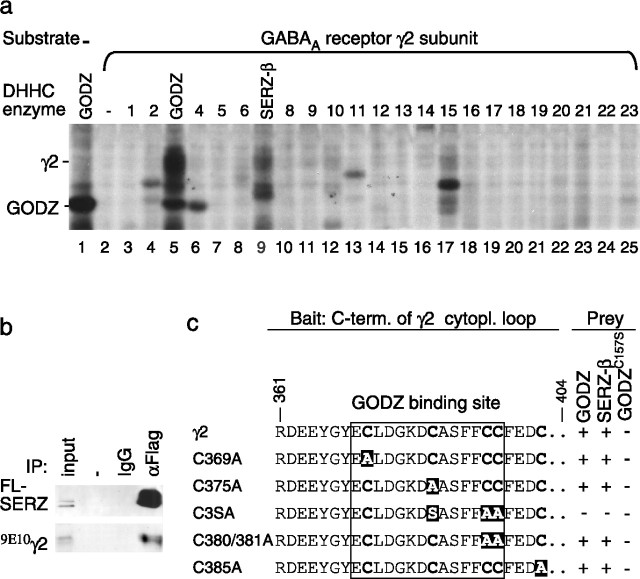
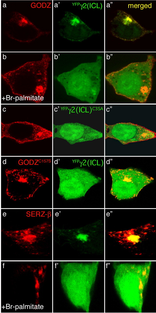
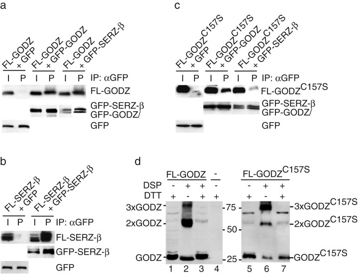
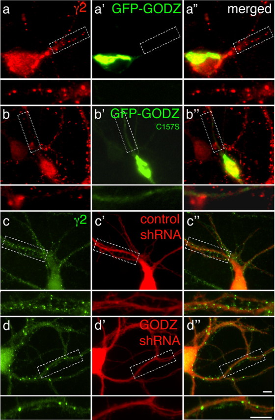
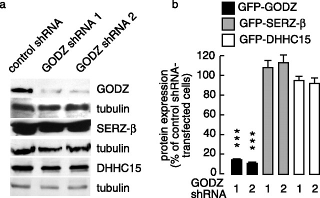
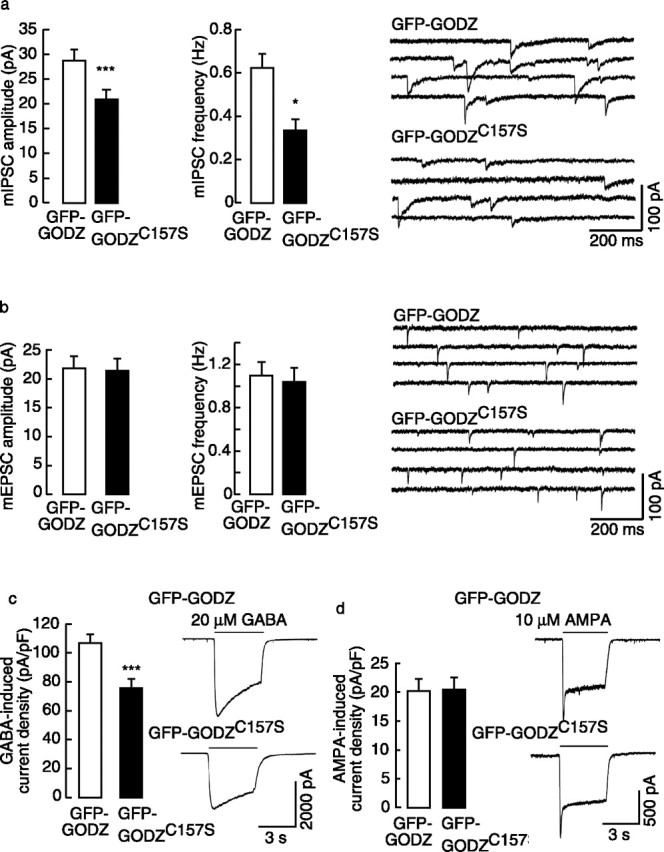
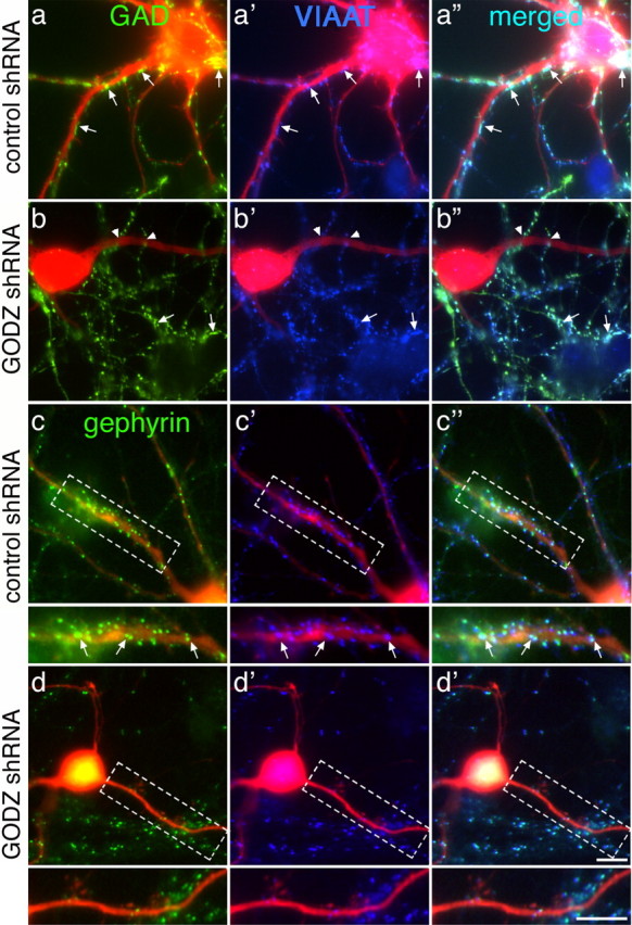
Golgi-specific DHHC (Asp-His-His-Cys) zinc finger protein (GODZ) is a DHHC family palmitoyl acyltransferase that is implicated in palmitoylation and regulated trafficking of diverse substrates that function either at inhibitory or excitatory synapses. Of particular interest is the gamma2 subunit of GABA(A) receptors, which is required for targeting these receptors to inhibitory synapses. Here, we report that GODZ and, to a lesser extent, its close paralog sertoli cell gene with a zinc finger domain-beta (SERZ-beta) are the main members of the DHHC family of enzymes that are able to palmitoylate the gamma2 subunit in heterologous cells. Yeast two-hybrid and colocalization assays in human embryonic kidney 293T (HEK293T) cells indicate that GODZ and SERZ-beta show indistinguishable palmitoylation-dependent interaction with the gamma2 subunit. After coexpression in HEK293T cells, they form homomultimers and heteromultimers, as shown by coimmunoprecipitation and in vivo cross-linking experiments. Analyses in neurons transfected with dominant-negative GODZ (GODZ(C157S)) or plasmid-based GODZ-specific RNAi indicate that GODZ is required for normal accumulation of GABA(A) receptors at synapses, for normal whole-cell and synaptic GABAergic inhibitory function and, indirectly, for GABAergic innervation. Unexpectedly, GODZ was found to be dispensable for normal postsynaptic AMPA receptor-mediated glutamatergic transmission. We conclude that GODZ-mediated palmitoylation of GABA(A) receptors and possibly other substrates contributes selectively to the formation and normal function of GABAergic inhibitory synapses.


Figures

Figure 1.
Figure 1.
GODZ and SERZ-β palmitoylate the γ2 subunit in vitro and interact with indistinguishable sequence specificity. a, Individual DHHC clones (1–23, with DHHC3 and 7 indicated as GODZ and SERZ-β, respectively) were cotransfected with (9E10)γ2 into HEK293T cells. The cells were metabolically labeled with 3H-palmitate and the proteins separated by SDS-PAGE and analyzed by fluorography. Note the selective labeling of the γ2 subunit in cells expressing GODZ (lane 5) or SERZ-β (lane 9), with a trace of labeling also apparent with DHHC15 (lane 17). No labeling above background was evident in the absence of transfected γ2 subunit (lane 1) or in the presence of any of the other DHHC proteins (lanes 2–4, 6–8, 10–25). b, HEK293T cells were cotransfected with the γ2 subunit and FL-SERZ-β and tested for interaction by coimmunoprecipitation and Western blot analysis. Precipitation of FL-SERZ-β was confirmed with mAb anti-FLAG. FLAG rabbit antiserum, but not IgG, resulted in efficient coimmunoprecipitation of the (9E10)γ2 subunit analogous to results previously shown for GODZ (Keller et al., 2004). c, Yeast two-hybrid assays using a γ2 subunit fragment (amino acids 361–404) or the indicated mutant derivatives as bait were used to test for interaction with full-length GODZ, SERZ-β, or GODZC157S as prey. The GODZ binding site (Keller et al., 2004) is boxed with the mutated cysteines highlighted in white on a black background. Note the identical sequence specificity of GODZ and SERZ-β for interaction with the γ2 subunit. Enzymatically inactive GODZC157S failed to interact with the γ2 constructs.
Figure 2.
Figure 2.
Palmitoylation-dependent interaction of GODZ and SERZ-β with the γ2 subunit. The DHHC proteins FL-GODZ (ac), FL-GODZC157S (d), or FL-SERZ-β (e, f) were cotransfected with YFPγ2(ICL) (a′, b′, d′f′) or its mutant derivative YFPγ2(ICL)C3SA (c′) and tested for colocalization after treatment of transfected cells with ethanol (aa″, ee″) or 2-BrP in ethanol (bb″, ff″), or without additional treatment (cc″, dd″). Immunofluorescently labeled FL-GODZ and FL-SERZ-β are shown in red (af), the YFP-tagged γ2 constructs are shown in green (a′f′), and colocalization of FL-GODZ or FL-SERZ-β with YFPγ2(ICL) is shown in yellow (a″f″). Restriction of YFPγ2(ICL) to the Golgi is indicative of interaction with GODZ or SERZ-β. Note the diffuse distribution of YFP-tagged γ2 constructs in experiments involving transfection of GODZC157S (d) or YFPγ2(ICL)C3SA (c) or treatment with 2-BrP (b, f), respectively, indicating that palmitoylation is required for interaction between substrate and PATs.
Figure 3.
Figure 3.
GODZ and SERZ-β form homomultimers and heteromultimers. a, FL-GODZ was cotransfected with GFP, GFP-GODZ, or GFP-SERZ-β into HEK293T cells. Aliquots of cell extracts (I, input) and αGFP immunoprecipitates (P) were analyzed by Western blot for coimmunoprecipitation of FL-GODZ with αFlag antiserum. The expression and immunoprecipitation of GFP proteins by αGFP antibody was verified on the same blot with rabbit αGFP antiserum. b, FL-SERZ-β was analyzed for coimmunoprecipitation with GFP-SERZ-β, as indicated in a. c, FL-GODZC157S was analyzed for coimmunoprecipitation with GFP-GODZ or GFP-SERZ-β, respectively, as indicated in a. d, FL-GODZ (left) or GODZC157S (right) were transfected into HEK293T cells and aliquots of the intact cells mock-treated or cross-linked with DSP. The cell extracts were processed for SDS-PAGE using loading buffer containing or lacking DTT (200 mm) and analyzed by Western blot using αFLAG antibody. Untransfected cells were processed as a control (lane 4). Note the DTT-sensitive cross-linking products corresponding in size to dimers and trimers of GODZ (lane 2, 3) and GODZC157S (lane 6, 7), respectively.
Figure 4.
Figure 4.
Overexpression of dominant-negative GODZ and GODZ-specific shRNA interferes with postsynaptic accumulation of GABAA receptors. a, b, Low-density cultures of cortical neurons (18 DIV) were transfected with GFP-GODZ (aa″) or GFP-GODZC157S (bb″) and subjected to immunofluorescent staining for the γ2 subunit 2 d later. Note the prominent punctate γ2 subunit staining in the GFP-GODZ-transfected neuron (a, red) and the highly restricted localization of GFP-GODZ to the Golgi complex (a′, green). In contrast, punctate immunoreactivity for the γ2 subunit was significantly reduced in the GFP-GODZC157S-transfected neuron (b). GFP-GODZC157S (b′) is concentrated in the Golgi complex and, unlike GFP-GODZ, also evident in dendrites. c, d, Cortical neurons were transfected with plasmid vectors encoding dsRed (red) and either control shRNA or GODZ-specific shRNA and processed for immunofluorescent analysis of the γ2 subunit as above. Note the significant reduction in punctate staining for the γ2 subunit (green) in the GODZ-shRNA-transfected neuron (d), compared with the neuron transfected with control shRNA (c). Merged images are shown in a″d″, with boxed dendritic segments shown enlarged in separate panels below each image. Scale bars, 5 μm.
Figure 5.
Figure 5.
Selective knock-down of GODZ by GODZ-specific shRNA. a, Analysis of the target specificity of shRNA constructs. A control vector encoding a mutated GODZ shRNA target sequence or one of two GODZ-specific shRNA constructs (GODZ shRNA 1, 2) were cotransfected with either FL-GODZ or FL-SERZ-β or FL-DHHC15 into HEK293T cells, and the cell extracts were analyzed by Western blot using mAb anti-FLAG. The blots were stripped and reprobed with anti-tubulin antibody as a loading control. Note the dramatic reduction in GODZ in cells cotransfected with GODZ shRNA 1 or 2, whereas SERZ-β and DHHC15 expression was unaffected under the same conditions. No effect on GODZ, SERZ-β, or DHHC15 expression was observed on cotransfection with the mutant shRNA control vector. b, Quantitation of GODZ shRNA-mediated effects on GODZ, SERZ-β, and DHHC15 expression in transfected HEK293T cells by flow cytometry (see Materials and Methods). The cells were transfected with GFP-GODZ, GFP-SERZ-β, or GFP-DHHC15 together with GODZ shRNA 1, GODZ shRNA 2, or control shRNA vector. Note the drastic reduction in GFP-GODZ expression by cotransfected GODZ shRNA1 (15.15 ± 0.91% of control shRNA; n = 6; p < 0.001) or GODZ shRNA2 (11.48 ± 1.48% of control; n = 6; p < 0.001). In contrast, neither of the two GODZ shRNA constructs had an effect on expression of GFP-SERZ-β (GODZ shRNA1, 109 ± 8.3% of control shRNA, n = 12, p > 0.05; GODZ shRNA2, 113 ± 8.7%, n = 12, p > 0.05) or GFP-DHHC15 (GODZ shRNA1, 95 ± 3.7% of control shRNA, n = 5, p > 0.05; GODZ shRNA2, 92 ± 5.0%, n = 6, p > 0.05).
Figure 6.
Figure 6.
Analyses of functional consequences of dominant-negative GODZ on GABAergic and glutamatergic transmission. GABAergic and glutamatergic miniature postsynaptic currents as well as GABA- and AMPA-induced whole-cell currents were recorded from transfected hypothalamic neurons (12–15 DIV). a, Analyses of mIPSCs of neurons transfected with dominant-negative GFP-GODZC157S revealed a significant reduction in both the amplitude (20.3 ± 1.4 pA) and frequency (0.31 ± 0.08 Hz; n = 26) of mIPSCs compared with matched GFP-GODZ-transfected controls (29.7 ± 2.0 pA, p < 0.001; 0.63 ± 0.10 Hz, n = 25, p < 0.05). b, In contrast, the mEPSC amplitude (21.9 ± 0.8 pA) and frequency (1.00 ± 0.24 Hz; n = 18) of GFP-GODZC157S-transfected neurons were indistinguishable from GFP-GODZ controls (22.4 ± 1.8 pA, 1.13 ± 0.24 Hz; n = 15; p > 0.05). c, The GABA (20 μm)-induced whole-cell current density in GODZC157S-transfected neurons (75.7 ± 4.4 pA/pF; n = 17) was significantly reduced compared with GFP-GODZ controls (107.0 ± 5.1 pA/pF; n = 14; p < 0.001). d, The AMPA (10 μm)-induced current density was unchanged in GFP-GODZC157S transfected neurons (19.1 ± 2.2 pA/pF; n = 15) compared with GFP-GODZ controls (19.8 ± 2.4 pA/F; n = 17; p > 0.05). Representative current traces are shown on the right of the respective bar diagrams. Data represent means ± SE (*p < 0.05, **p < 0.01, ***p < 0.001, Student's t test).
Figure 7.
Figure 7.
GODZ shRNA expression in postsynaptic neurons interferes with GABAergic innervation. Cortical neurons were transfected with either control shRNA (aa″, cc″) or GODZ shRNA (bb″, dd″) as described in Figure 5 and double-immunostained for GAD (a, b, green) or gephyrin (c, d, green) together with VIAAT (a′, b′, c′, d′, blue), with merged images shown in a″, b″, c″, and d″. Transfected neurons and their major processes are identified by the red fluorescence of shRNA vector-encoded dsRed (red). Note the GAD- and VIAAT-positive GABAergic varicosities (arrows) that are evident along most of the dendrites of a representative control shRNA-transfected neuron (aa″). In contrast, the GODZ shRNA-transfected neuron (bb″) is essentially devoid of contact with GABAergic axons, which, if present, appear to grow across (arrowheads) rather than along dendrites. A nearby, untransfected neuron in the same image (bb″) is highly innervated (arrows), similar to the control shRNA-transfected neuron shown in aa″. The punctate staining for gephyrin in a control shRNA transfected neuron (c) is perfectly colocalized with immunofluorescent staining of presynaptic VIAAT (c′, c″, arrows), and staining for both of these marker proteins is greatly reduced in a GODZ shRNA-transfected neuron (d, d′, d″). Note the untransfected pair of dendrites in the same image that is decorated with gephyrin clusters and strongly innervated by GABAergic axons. Scale bars, 5 μm.

Similar articles

Cited by

References

    1. Alldred MJ, Mulder-Rosi J, Lingenfelter SE, Chen G, Luscher B. Distinct γ2 subunit domains mediate clustering and synaptic function of postsynaptic GABAA receptors and gephyrin. J Neurosci. 2005;25:594–603. - PMC - PubMed
    1. Baer K, Essrich C, Benson JA, Benke D, Bluethmann H, Fritschy J-M, Luscher B. Postsynaptic clustering of GABAA receptors by the γ3 subunit in vivo. Proc Natl Acad Sci USA. 1999;96:12860–12865. - PMC - PubMed
    1. Bedford FK, Kittler JT, Muller E, Thomas P, Uren JM, Merlo D, Wisden W, Triller A, Smart TG, Moss SJ. GABAA receptor cell surface number and subunit stability are regulated by the ubiquitin-like protein Plic-1. Nat Neurosci. 2001;4:908–916. - PubMed
    1. Bijlmakers MJ, Marsh M. The on-off story of protein palmitoylation. Trends Cell Biol. 2003;13:32–42. - PubMed
    1. Bogdanov Y, Michels G, Armstrong-Gold C, Haydon PG, Lindstrom J, Pangalos M, Moss SJ. Synaptic GABA(A) receptors are directly recruited from their extrasynaptic counterparts. EMBO J. 2006;25:4381–4389. - PMC - PubMed

Publication types

MeSH terms