

Effect of the conceptus on uterine natural killer cell numbers and function in the mouse uterus during decidualization
- PMID: 17151350
- PMCID: PMC2275212
- DOI: 10.1095/biolreprod.106.056630
Effect of the conceptus on uterine natural killer cell numbers and function in the mouse uterus during decidualization
Abstract
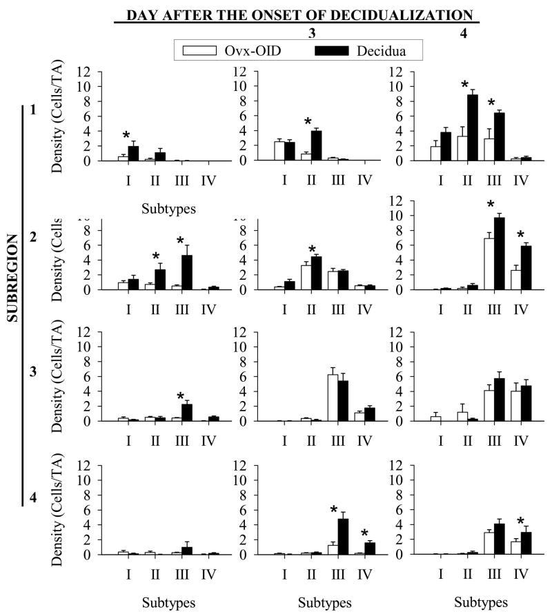
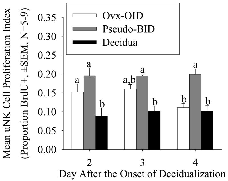
Uterine natural killer (uNK) cells are the most abundant lymphocytes in the uterus during early pregnancy and play a role in spiral arteriole modifications. In the present study, we investigated whether uNK cell populations differed between mouse decidua and deciduoma. Histochemical staining using the Dolichos biflorus agglutinin (DBA) lectin was used to identify uNK cells and classify their stages of maturation. We found differences in the pattern of localization and density of uNK cells between the decidua and deciduoma at Days 2-4 after the onset of decidualization. The cells were more distributed and the densities were significantly greater in the mesometrial region of the decidua than in the deciduoma. Using double-labeling for DBA lectin binding and bromodeoxyuridine incorporation, we found that the higher number of uNK cells in the decidua was not due to an increase in uNK cell proliferation. Western blot analyses revealed that the increase in uNK cell number was accompanied by significant increases in the levels of interferon gamma (IFNG) and prointerleukin 18 when a conceptus was present. Vascular morphometry revealed that modifications of the spiral arterioles occurred in the mesometrial decidua but not in the deciduoma, which could be attributed to the differences observed in uNK cell number and IFNG production. The present study demonstrates that differences exist in uNK cell populations between the decidua and deciduoma, providing evidence that the conceptus generates signals that regulate uNK cell number and function in the uterus during implantation.
Figures


Similar articles
-
The conceptus increases secreted phosphoprotein 1 gene expression in the mouse uterus during the progression of decidualization mainly due to its effects on uterine natural killer cells.Reproduction. 2007 Jun;133(6):1213-21. doi: 10.1530/REP-07-0085. Reproduction. 2007. PMID: 17636175 Free PMC article.
-
A map of relationships between uterine natural killer cells and progesterone receptor expressing cells during mouse pregnancy.Placenta. 2008 Apr;29(4):317-23. doi: 10.1016/j.placenta.2008.01.003. Epub 2008 Mar 4. Placenta. 2008. PMID: 18289671
-
Splenectomy delays uterine natural killer cell recruitment to implantation sites and prolongs pregnancy in mice.Anat Rec (Hoboken). 2012 Jul;295(7):1221-8. doi: 10.1002/ar.22498. Epub 2012 May 17. Anat Rec (Hoboken). 2012. PMID: 22593087
-
The regulation of ovary and conceptus on the uterine natural killer cells during early pregnancy.Reprod Biol Endocrinol. 2017 Sep 6;15(1):73. doi: 10.1186/s12958-017-0290-1. Reprod Biol Endocrinol. 2017. PMID: 28874155 Free PMC article. Review.
-
Uterine natural killer cells: insights into their cellular and molecular biology from mouse modelling.Reproduction. 2003 Aug;126(2):149-60. doi: 10.1530/rep.0.1260149. Reproduction. 2003. PMID: 12887272 Free PMC article. Review.
Cited by
-
Leukocyte driven-decidual angiogenesis in early pregnancy.Cell Mol Immunol. 2014 Nov;11(6):522-37. doi: 10.1038/cmi.2014.63. Epub 2014 Jul 28. Cell Mol Immunol. 2014. PMID: 25066422 Free PMC article. Review.
-
Physiological and molecular determinants of embryo implantation.Mol Aspects Med. 2013 Oct;34(5):939-80. doi: 10.1016/j.mam.2012.12.011. Epub 2013 Jan 2. Mol Aspects Med. 2013. PMID: 23290997 Free PMC article. Review.
-
Immunomorphological changes in the rhesus monkey endometrium and decidua during the menstrual cycle and early pregnancy.Am J Reprod Immunol. 2012 Oct;68(4):309-21. doi: 10.1111/j.1600-0897.2012.01174.x. Epub 2012 Jul 11. Am J Reprod Immunol. 2012. PMID: 22784010 Free PMC article.
-
The conceptus increases secreted phosphoprotein 1 gene expression in the mouse uterus during the progression of decidualization mainly due to its effects on uterine natural killer cells.Reproduction. 2007 Jun;133(6):1213-21. doi: 10.1530/REP-07-0085. Reproduction. 2007. PMID: 17636175 Free PMC article.
-
Tumor necrosis factor receptor subfamily 9 (Tnfrsf9) gene is expressed in distinct cell populations in mouse uterus and conceptus during implantation period of pregnancy.Cell Tissue Res. 2011 Jun;344(3):567-76. doi: 10.1007/s00441-011-1171-0. Epub 2011 May 12. Cell Tissue Res. 2011. PMID: 21560035 Free PMC article.
References
-
- Das RM, Martin L. Uterine DNA synthesis and cell proliferation during early decidualization induced by oil in mice. J Reprod Fertil. 1978;53:125–128. - PubMed
-
- Lejeune B, Van Hoeck J, Leroy F. Satellite versus total DNA replication in relation to endopolyploidy of decidual cells in the mouse. Chromosoma. 1982;84:511–516. - PubMed
-
- Loeb L. The production of deciduomata and the relation between the ovaries and the formation of the decidua. J Am Med Assoc. 1908;50:1897–1901.
-
- Finn CA, Martin L. The control of implantation. J Reprod Fertil. 1974;39:195–206. - PubMed
-
- Krehbiel RH. Cytological studies of the decidual reaction in the rat during early pregnancy and in the production of the deciduomata. Physiological Zoology. 1937;X:212–233.
Publication types
MeSH terms
Substances
Grants and funding
LinkOut - more resources
Full Text Sources
