. 2007 Feb;18(2):547-56.
doi: 10.1091/mbc.e06-09-0776. Epub 2006 Dec 6.

The plasma membrane proteins Prm1 and Fig1 ascertain fidelity of membrane fusion during yeast mating

Affiliations

The plasma membrane proteins Prm1 and Fig1 ascertain fidelity of membrane fusion during yeast mating

Pablo S Aguilar et al. Mol Biol Cell. 2007 Feb.

Abstract

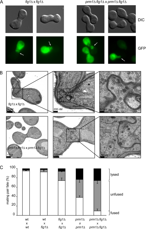
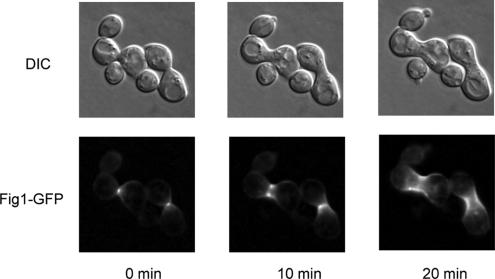
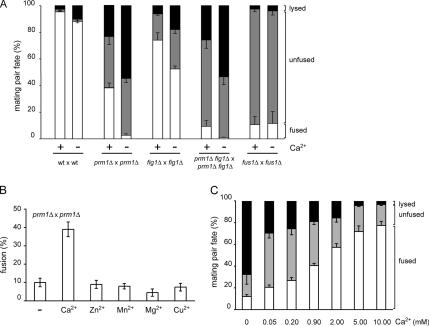
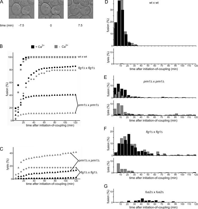
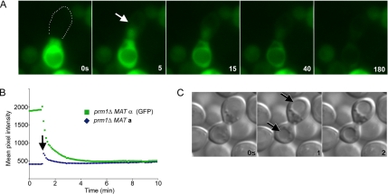
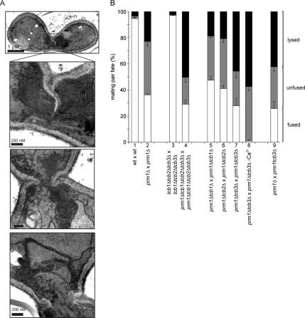
As for most cell-cell fusion events, the molecular details of membrane fusion during yeast mating are poorly understood. The multipass membrane protein Prm1 is the only known component that acts at the step of bilayer fusion. In its absence, mutant mating pairs lyse or arrest in the mating reaction with tightly apposed plasma membranes. We show that deletion of FIG 1, which controls pheromone-induced Ca(2+) influx, yields similar cell fusion defects. Although extracellular Ca(2+) is not required for efficient cell fusion of wild-type cells, cell fusion in prm1 mutant mating pairs is dramatically reduced when Ca(2+) is removed. This enhanced fusion defect is due to lysis. Time-lapse microscopy reveals that fusion and lysis events initiate with identical kinetics, suggesting that both outcomes result from engagement of the fusion machinery. The yeast synaptotagmin orthologue and Ca(2+) binding protein Tcb3 has a role in reducing lysis of prm1 mutants, which opens the possibility that the observed role of Ca(2+) is to engage a wound repair mechanism. Thus, our results suggest that Prm1 and Fig1 have a role in enhancing membrane fusion and maintaining its fidelity. Their absence results in frequent mating pair lysis, which is counteracted by Ca(2+)-dependent membrane repair.


Figures

Figure 1.
Figure 1.
PRM1 and FIG 1 promote the membrane fusion step during mating. (A) Unfused fig1Δ × fig1Δ mating pairs form bubbles. MATα cells carrying cytoplasmic GFP were mixed with MATa cells on nitrocellulose filters and incubated on YPD plates for 3 h at 30°C. Fixed mating mixtures were then imaged by DIC and fluorescence microscopy. Arrows point to mating pair bubbles. (B) Bubbles in unfused fig1Δ × fig1Δ mating pairs contain closely apposed membranes. The ultrastructural detail of fig1Δ × fig1Δ mating pairs was determined as described in A with mating mixtures processed as described in Materials and Methods. The panels show three different magnifications for each mating pair. The lower magnification picture in the bottom panel corresponds to a different section of the same mating pair. (C) PRM1 and FIG 1 act through different pathways to promote cell fusion. Quantitative cell fusion and lysis assays were performed as described in Materials and Methods. All deletion genotypes were tested for both mating types; the results were indistinguishable. Error bars indicate SEs for four independent experiments with 300 mating pairs scored for each case.
Figure 2.
Figure 2.
Fig1 localizes at the site of cell fusion. Cells of opposite mating types bearing the FIG 1–GFP fusion gene were mixed, preincubated on filters for 1 h on YPD at 30°C, and mounted on agarose complete media pads for imaging.
Figure 3.
Figure 3.
Extracellular Ca2+ suppresses cell lysis in prm1Δ × prm1Δ mating reactions. (A) Fusion of prm1Δ × prm1Δ mating pairs is highly sensitive to EGTA. Mating mixtures were incubated on YPD plates with (Ca2+, −) or without (Ca2+, +) 20 mM EGTA. (B) Sensitivity of prm1Δ × prm1Δ mating pairs to EGTA is due to a lack of Ca2+. Liquid media cell fusion assays were performed in synthetic media treated with resin-bound BAPTA and supplemented either with water (−) or divalent cation salts at concentrations found in synthetic complete media: 900 μM calcium chloride, 1 μM zinc sulfate, 10 μM manganese sulfate, 1 mM magnesium sulfate, and 1 μM copper sulfate. (C) Extracellular calcium suppresses the prm1Δ × prm1Δ mating defect. Liquid media cell fusion assays were performed as in B by using media supplemented with calcium chloride as indicated. Error bars indicate SEs for at least three independent experiments with 300 mating pairs scored per experiment.
Figure 4.
Figure 4.
Kinetics of fusion and lysis initiation in mating populations. The fate of individual mating pairs in a mating mixture was followed by time-lapse microscopy. (A) Example of the determination of initiation of coupling. The picture in the middle, showing the two cells initiating contact in a polarized manner, serves as a time-zero reference point for the mating pair. (B) Plot showing the progression of the fusion outcome as a function of time in the presence (black symbols) or absence (gray symbols) of Ca2+. (C) Plot showing the progression of the lysis outcome as a function of time. (D–G) Fusion and lysis events were binned into 5-min time windows and normalized to indicate the percentage of the mating pair population that would undergo the indicated event within these time windows. Black bars represent events in the presence of Ca2+; gray bars represent events in the absence of Ca2+. All data shown are the result of three independent experiments. No lysis was observed for fus2Δ × fus2Δ. The lower rate of lysis (compared with the endpoint assays shown in Figures 1 and 2) is likely due to performing the mating reaction at room temperature instead of at 30°C.
Figure 5.
Figure 5.
Lysis occurs simultaneously in each cell of a mating pair and is concomitant with cytoplasmic mixing. Lysis of prm1Δ × prm1Δ mating pairs in media lacking Ca2+ was imaged with fast time resolution. (A) Cytoplasmic GFP in a MATα cell spread into the MATa cell after initiation of lysis at 5 s (white arrow). Note that GFP has spread throughout the entire MATa cell in all frames subsequent to the 0-s frame, demonstrating cytoplasmic continuity. White dots represent the boundaries of the MATa cell. (B) Quantification of average pixel intensity in each mating partner over time; initiation of lysis is indicated by the black arrow. (C) DIC images of a lysing mating pair. Evidence of lysis is seen in the 1 s frame as rounded vacuoles (black arrows).
Figure 6.
Figure 6.
TCB3 prevents mating pairs lysis in the absence of PRM1. (A) In the absence of Ca2+, prm1Δ × prm1Δ mating pairs show extensive membrane accumulation in the zone of cell fusion. Mating mixtures were processed as described in Materials and Methods. The panels show the zone of cell–cell contact for three different mating pairs. (B) Deletions of TCB1, TCB2, and TCB3 were tested for their effect on cell fusion and lysis during mating. Crosses are labeled as MATα × MATa. Error bars indicate SEs for at least three independent experiments with 300 mating pairs scored per experiment.

References

    1. Bagnat M., Simons K. Cell surface polarization during yeast mating. Proc. Natl. Acad. Sci. USA. 2002;99:14183–14188. - PMC - PubMed
    1. Chen E. H., Olson E. N. Unveiling the mechanisms of cell-cell fusion. Science. 2005;308:369–373. - PubMed
    1. Creutz C. E., Snyder S. L., Schulz T. A. Characterization of the yeast tricalbins: membrane-bound multi-C2-domain proteins that form complexes involved in membrane trafficking. Cell Mol. Life Sci. 2004;61:1208–1220. - PMC - PubMed
    1. Duzgunes N., Wilschut J., Fraley R., Papahadjopoulos D. Studies on the mechanism of membrane fusion. Role of head-group composition in calcium- and magnesium-induced fusion of mixed phospholipid vesicles. Biochim. Biophys. Acta. 1981;642:182–195. - PubMed
    1. Ellens H., Bentz J., Szoka F. C. H+- and Ca2+-induced fusion and destabilization of liposomes. Biochemistry. 1985;24:3099–3106. - PubMed

Publication types