. 2007 Jan 1;195(1):37-45.
doi: 10.1086/509823. Epub 2006 Nov 20.

Vaccinia virus inhibits T cell receptor-dependent responses by human gammadelta T cells

Affiliations

Vaccinia virus inhibits T cell receptor-dependent responses by human gammadelta T cells

Haishan Li et al. J Infect Dis. .

Abstract

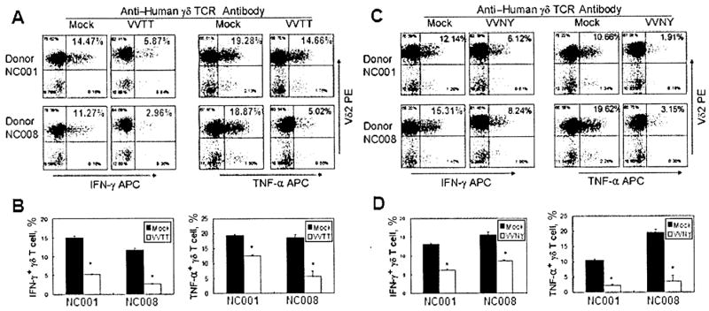
Vaccinia virus (VV) is an effective vaccine and vector but has evolved multiple mechanisms for evading host immunity. We characterized the interactions of VV (TianTan and New York City Board of Health strains) with human gammadelta T cells because of the role they play in immune control of this virus. Exposure to VV failed to trigger proliferative responses in gammadelta T cells from unprimed individuals, but it was an unexpected finding that VV blocked responses to model antigens by the Vgamma2Vdelta2 T cell subset. Infectious or ultraviolet light-inactivated VV inhibited proliferative Vgamma2Vdelta2 T cell responses to phosphoantigens and tumor cells, prevented cytolysis of Daudi B cells, and reduced cytokine production. Inhibiting Vgamma2Vdelta2 T cells may be a mechanism for evading host immunity and increasing VV virulence. Increased VV replication or expression in the absence of gammadelta T cell responses might contribute to its potency as a vaccine against poxvirus and recombinant antigens.


Conflict of interest statement

Potential conflicts of interest: none reported.

Figures

Figure 1
Figure 1
Lack of stimulation of γδ T cell expansion in vitro by vaccinia virus (VV). Peripheral-blood mononuclear cells (PBMCs) from 2 healthy, unrelated donors were stained for PANγδ and CD3 on day 0 and 14 days after stimulation with isopentyl pyrophosphate (IPP), UV light–inactivated TianTan strain carrying the HIV gag gene (UV-VVTT; MOI of 1, 0.1, or 0.01), or irradiated VV-infected CEM cells (CEM/VVTT; CEM/VVTT:PBMC ratio of 1, 0.1, or 0.01). Although IPP stimulated obvious expansion of γδ T cells, UV-VVTT and CEM/VVTT did not induce any expansion of γδ T cells. APC, allophycocyanin; FITC, fluorescein isothiocyanate.
Figure 2
Figure 2
Inhibition of isopentyl pyrophosphate (IPP)– or Daudi cell–stimulated expansion of human γδ T cells by vaccinia virus (VV). A, Dose dependency of inhibition. UV light–inactivated VV (UV-VV; either the TianTan strain carrying the HIV gag gene [VVTT] or the nonrecombinant New York City Board of Health strain [VVNY]; MOI of 1, 0.1, or 0.01), irradiated VVTT- or VVNY-infected CEM cells (CEM/VV; CEM/VV:peripheral-blood mononuclear cell [PBMC] ratio of 1:1, 1:10, or 1:100), or appropriate controls were added to IPP/interleukin (IL)–2–treated cultures. On day 14, the expansion was measured by flow cytometry. IPP-driven expansion of γδ T cells was inhibited by both UV-VV and CEM/VV in a dose-dependent manner, compared with that in mock-treated or CEM cell controls. B, Repetition of experiment in 3 additional donors. At an MOI of 1 for UV-VVTT or a CEM/VVTT:PBMC ratio of 1:1, we obtained similar results for all donors, showing complete suppression of Vγ2Vδ2 T cell proliferation. An effect of VV on Daudi cell–stimulated expansion of γδ T cells was also detected. UV-VVTT (MOI of 1) was added to Daudi/IL-2–treated cultures or Daudi B cells were preinfected with VV (MOI of 1) for 20 h before being added to the culture system. The results indicated that Daudi cell–stimulated expansion of γδ T cells was completely inhibited. C, Little inhibitory effect of VVTT on phytohemagglutinin (PHA)–stimulated proliferation of CD4+ or CD43 cells in PBMCs. PBMCs were stained with carboxyfluorescein diacetate succinimidyl ester (CFSE), cultured in 96-well round-bottomed plates in the presence of either UV-VV (VVTT or VVNY) at an MOI of 1 or CEM/VV (VVTT or VVNY) at a CEM/VV:PBMC ratio of 1:1, stimulated with 5 μg/mL PHA and 100 U/mL IL-2 for 4 days, and stained with anti-CD4 antibody for the flow cytometry assay. APC, allophycocyanin; FITC, fluorescein isothiocyanate.
Figure 3
Figure 3
Alteration of the length distribution of Vγ2 chains or reduction in viability not induced by vaccinia virus (VV) in a Vγ2Vδ2 T cell line. Total RNA was extracted from cells before stimulation or 14 days after stimulation with isopentyl pyrophosphate (IPP), IPP plus irradiated CEM cells infected with the TianTan VV strain carrying the HIV gag gene (CEM/VVTT; CEM/VVTT:peripheral-blood mononuclear cell [PBMC] ratio of 1:1), UV light–inactivated VVTT (UV-VVTT; MOI of 1), or live VVTT (MOI of 1) (A). cDNA was synthesized, and a spectratype assay was performed. The proportion of Vγ2-Jγ1.2 chains was not significantly changed after VV exposure (B). A Vγ2Vδ2 T cell line was generated by IPP stimulation of PBMCs and proliferation of the γδ T cell subset (C). These cells were exposed to VV and samples were collected for 35 h, to measure the frequency of Vγ2-Jγ1.2 chains. There were no significant changes over this time interval. The Vγ2Vδ2 T cell line was cultured with VVTT for 3 days without substantial loss of cell viability (D).
Figure 4
Figure 4
Inhibition of Daudi cell cytotoxicity of γδ T cells by vaccinia virus (VV). Isopentyl pyrophosphate (IPP)–expanded γδ T cells from several donors were incubated with purified TianTan strain carrying the HIV gag gene (VVTT) or UV light–inactivated VVTT (UV-VVTT) at an MOI of 1 or with mock control for 2 h, and then the ability of these cells to lyse Daudi B target cells was determined by calcein-release cytotoxicity assay. E:T ratio, effector-to-target cell ratio.
Figure 5
Figure 5
Reduction in cell-surface CD107a expression not induced by vaccinia virus (VV) after stimulation with anti–human γδ T cell receptor (TCR) antibody. Isopentyl pyrophosphate (IPP)–expanded γδ T cells from 2 donors were incubated with purified VVTT at an MOI of 1 as well as with mock control for 2 h and then stimulated with anti–human γδ TCR antibody for 2 h and stained for Vδ2 and CD107a. FITC, fluorescein isothiocyanate; PE, phycoerythrin.
Figure 6
Figure 6
Inhibition of γδ T cell interferon (IFN)–γ and tumor necrosis factor (TNF)–α production by vaccinia virus (VV) after stimulation with anti–human γδ T cell receptor (TCR) antibody. Isopentyl pyrophosphate (IPP)–expanded γδ T cells from 2 donors were incubated with purified VV (either the TianTan strain carrying the HIV gag gene [VVTT] or the nonrecombinant New York City Board of Health strain [VVNY]) at an MOI of 1 as well as with mock control for 2 h, stimulated with anti–human γδ TCR antibody for 2 h, and stained for Vδ2 and intracellular IFN-γ or TNF-α (A and C ). Intracellular staining for IFN-γ and TNF-α was performed in triplicate (B and D ). Statistically significant differences (P < .05) are indicated by asterisks. APC, allophycocyanin; PE, phycoerythrin.

Similar articles

Cited by

References

    1. Fenner F. History of international public health. 6. Geneva: World Health Organization; 1988. World Health Organization. Smallpox and its eradication.
    1. Moss B. Genetically engineered poxviruses for recombinant gene expression, vaccination, and safety. Proc Natl Acad Sci USA. 1996;93:11341–8. - PMC - PubMed
    1. Haga IR, Bowie AG. Evasion of innate immunity by vaccinia virus. Parasitology. 2005;130(Suppl):S11–25. - PubMed
    1. Seet BT, Johnston JB, Brunetti CR, et al. Poxviruses and immune evasion. Annu Rev Immunol. 2003;21:377–423. - PubMed
    1. Kirwan S, Merriam D, Barsby N, McKinnon A, Burshtyn DN. Vaccinia virus modulation of natural killer cell function by direct infection. Virology. 2006;347:75–87. - PubMed

Publication types

MeSH terms

Substances