. 2007 Jan;25(1):48-54.
doi: 10.1002/jmri.20810.

Two-dimensional population map of cortical connections in the human internal capsule

Affiliations

Two-dimensional population map of cortical connections in the human internal capsule

Mojtaba Zarei et al. J Magn Reson Imaging. 2007 Jan.

Abstract

Purpose: To exploit diffusion imaging tractography to produce a two-dimensional (xy) probabilistic population map of the cortical connections within the human internal capsule (IC).

Materials and methods: Diffusion tensor imaging (DTI) was carried out on 11 healthy volunteers. We parceled an axial section of the IC according to its connections to the prefrontal, premotor, primary motor, primary somatosensory, posterior parietal, and occipital cortices using our locally developed probabilistic algorithm.

Results: A consistent topographical organization was generated that was consistent with our anatomical knowledge of the IC. In addition, our study shows that in humans the anterior half of the IC is occupied by the prefrontal cortex.

Conclusion: This map may be of clinical use for correlating neurological deficit with hemispheric lesions, particularly those that affect sensorimotor function.


Figures

Figure 1
Figure 1
Group probability maps for each cortical region show a remarkable reproducibility of location of cortical connections within the IC across individuals. Prefrontal cortical connections occupied half of the entire volume of the IC. Sensorimotor tracts are in the posterior half of the PLIC. All cortical tracts are seen in both the lower (z = 1) and upper (z = 7) IC masks. Voxels are thresholded at 10% connection probability. Abbreviations: PFC = prefrontal, M1 = primary motor, S1 = primary somatosensory, PPC = posterior parietal, PMC = premotor, Occ = occipital, Tmp=temporal.
Figure 2
Figure 2
a: Hard segmentation map for the population, showing a topographical organization of cortical tracts in the IC that is very similar to the organization seen in the cerebral cortex. b: A group probability map that was thresholded to include voxels that were present in >30% of the group illustrates the overlap between connection regions, particularly the premotor cortex and M1, and M1 and S1. Abbreviations as in Fig. 1.
Figure 3
Figure 3
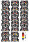
Hard segmentation maps for each individual studied, showing a remarkable reproducibility and consistency across subjects. They also demonstrate the consistency of topography of voxels at both axial levels. Voxels are thresholded at 10% connection probability. Abbreviations as in Fig. 1.
Figure 4
Figure 4
The IC tract-to-cortex volume ratio for the cortical connections in the IC shows that sensorimotor tracts make up the largest relative volume in the IC. This suggests that the sensorimotor connections have denser fiber tracts than other cortical tracts. In addition, this ratio is higher in the left hemisphere, which may suggest a correlation with the cerebral dominance. A repeated-measures analysis of variance (ANOVA) showed significant main effects of hemisphere (F = 18.3, P = 0.002) and cortical target (F = 43.6, P < 0.001), and a significant interaction between hemisphere and cortical target (F = 5.9, P < 0.001). On average, the seed to target volume percentile was greater for the left hemisphere, and this effect was significant by paired t-tests in the primary motor (t = -3.4, P = 0.007), premotor (t = -2.8, P = 0.017), and primary somatosensory (t = -3.0, P = 0.013) cortices. Abbreviations as in Fig. 1.

References

    1. Dejerine J, Déjerine-Klumpke A. Anatomie des centres nerveaux. Paris: Rueff; 1901. p. 816.
    1. Hardy TL, Bertrand G, Thompson CJ. The position and organization of motor fibers in the internal capsule found during stereotactic surgery. Appl Neurophysiol. 1979;42:160–170. - PubMed
    1. Hardy TL, Bertrand G, Thompson CJ. Organization and topography of sensory responses in the internal capsule and nucleus ventralis caudalis found during stereotactic surgery. Appl Neurophysiol. 1980;42:335–351. - PubMed
    1. Englander RN, Netsky MG, Adelman LS. Location of human pyramidal tract in the internal capsule: anatomic evidence. Neurology. 1975;25:823–826. - PubMed
    1. Leon SF, Pradilla AG. Antero-posterior localization of the pyramidal tract in the internal capsule in a living patient. Invest Clin. 1994;35:67–75. - PubMed