

Case Reports
Complications of Meckel's diverticula in adults
Affiliations
- PMID: 17152574
- PMCID: PMC3207587
Item in Clipboard
Case Reports
Complications of Meckel's diverticula in adults
Can J Surg.
2006 Oct.
No abstract available
Figures
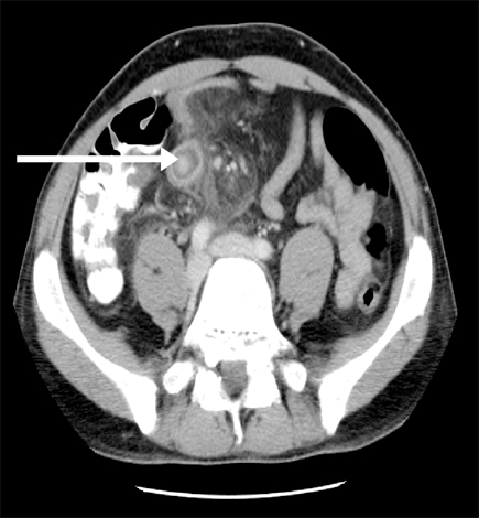
FIG. 1. CT abdomen/pelvis enhanced with oral and intravenous contrast. Target lesion (arrow) suggests terminal ileum wall thickening, while mesentery of the distal small bowel demonstrates mesenteric stranding.
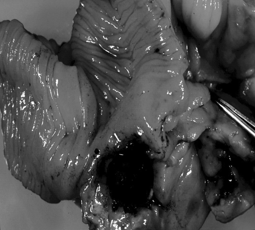
FIG. 2. A resection of distal small bowel. Approximately 5.0 cm from the proximal margin (34.0 cm from the terminal ileum) is an out-pouching of the small bowel that is occluded in the neck area by a fecalith measuring 1.5 cm. Distal to the fecalith (not shown) is a thick walled cavity lined with edematous bowel wall and firm fatty tissue.
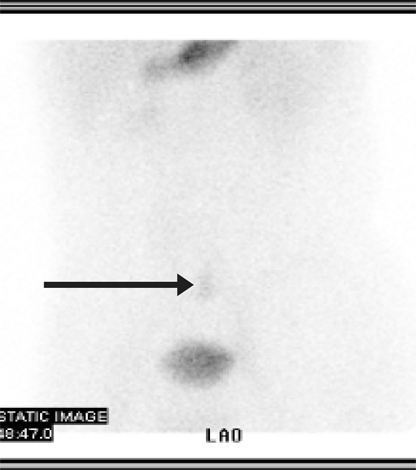
FIG. 3. Meckel's scan. A focus of abnormal radiotracer uptake is demonstrated in the lower abdomen several centimetres above the dome of the bladder. In this clinical situation the appearance is consistent with a Meckel's diverticulum containing ectopic gastric mucosa.
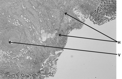
FIG. 4. Small bowel mucosa with hemorrhagic ulceration (H) and a feeder vessel (V).
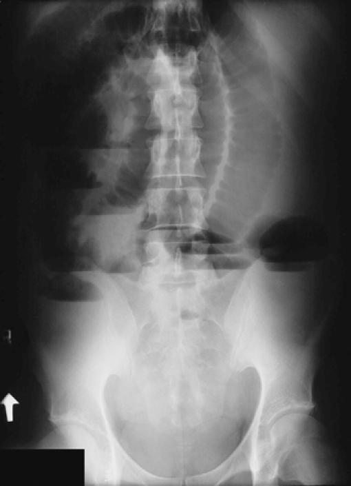
FIG. 5. Upright abdominal x-ray showing dilated loops of small bowel with multiple air fluid levels.
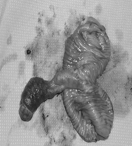
FIG. 6. Intussuscepted portion of small bowel exposing an inverted Meckel's diverticulum (3.3 cm in length). Projecting from the tip is a firm hemorrhagic mass with ulcerations. Sectioning of the mass reveals a lipoma (3.9 × 1.5 × 1.9 cm).
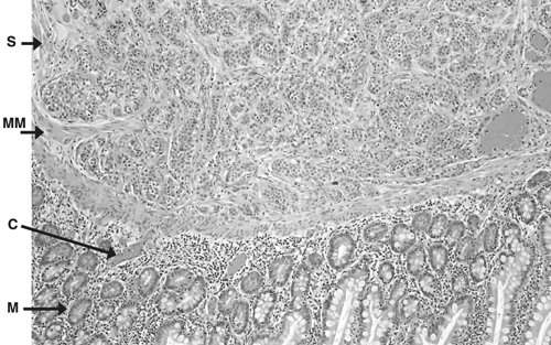
FIG. 7. Normal histology of the Meckel's diverticula, with small foci of carcinoid tumour. Normal mucosa (M) and normal submucosa (S), separated by muscularis mucosa (MM). Several small foci (< 0.2 cm each) of carcinoid tumour (C) are identified at the interface of the lamina propria and sub mucosa. The tumour does not invade the luminal surface or extend into the muscularis propria.
Comment in
-
Meckel's diverticulum causing mechanical small bowel obstruction.Can J Surg. 2008 Apr;51(2):156. Can J Surg. 2008. PMID: 18377760 Free PMC article. No abstract available.
References
-
- Meckel JF. Uber die divertikel am darmkanal. Arch Die Physio 1809;9:421-53.
-
- Kuwajerwala NK, Silva YJ. Meckel diverticulum. Available from: http://www.emedicine.com/med/topic2797.htm. Accessed March 2001.
-
- Stone PA, Hofeldt MJ, Campbell JE, et al. Meckel diverticulum: ten-year experience in adults. South Med J 2004;97:1038-41. - PubMed
-
- Turgeon DK, Barnett JL. Meckel diverticulum. Am J Gastroenterol 1990;85:777-81. - PubMed
-
- Yahchouchy EK, Marano AF, Etienne JCF, et al. Meckel's diverticulum. J Am Coll Surg 2001;192:658-62. - PubMed
Publication types
MeSH terms
LinkOut - more resources
Full Text Sources
Medical