. 2006 Dec 19;45(50):15020-8.
doi: 10.1021/bi061566o.

Importance of force linkage in mechanochemistry of adhesion receptors

Affiliations

Importance of force linkage in mechanochemistry of adhesion receptors

Nathan S Astrof et al. Biochemistry. .

Abstract

The alpha subunit-inserted (I) domain of integrin alphaLbeta2 [lymphocyte function-associated antigen-1 (LFA-1)] binds to intercellular adhesion molecule-1 (ICAM-1). The C- and N-termini of the alpha I domain are near one another on the "lower" face, opposite the metal ion-dependent adhesion site (MIDAS) on the "upper face". In conversion to the open alpha I domain conformation, a 7 A downward, axial displacement of C-terminal helix alpha7 is allosterically linked to rearrangement of the MIDAS into its high-affinity conformation. Here, we test the hypothesis that when an applied force is appropriately linked to conformational change, the conformational change can stabilize adhesive interactions that resist the applied force. Integrin alpha I domains were anchored to the cell surface through their C- or N-termini using type I or II transmembrane domains, respectively. C-terminal but not N-terminal anchorage robustly supported cell rolling on ICAM-1 substrates in shear flow. In contrast, when the alphaL I domain was mutationally stabilized in the open conformation with a disulfide bond, it mediated comparable levels of firm adhesion with type I and type II membrane anchors. To exclude other effects as the source of differential adhesion, these results were replicated using alpha I domains conjugated through the N- or C-terminus to polystyrene microspheres. Our results demonstrate a mechanical feedback system for regulating the strength of an adhesive bond. A review of crystal structures of integrin alpha and beta subunit I domains and selectins in high- and low-affinity conformations demonstrates a common mechanochemical design in which biologically applied tensile force stabilizes the more extended, high-affinity conformation.


Figures

Figure 1
Figure 1
I domain allostery and force. (A) Schematic representation of the I domain. The transition of the MIDAS from the closed to open conformation is linked to an axial, C-terminal shift of the C-terminal α helix (arrow). (B) Schematic of a cell adhering to a substrate in shear flow, with hydrodynamic force on the cell (FS) and force on the tether (FT). (C and D) The boxed region in panel B is magnified to show the tether to the substrate through the I domain–ICAM-1 interaction. ICAM-1 is shown linked to the substrate (straight line, bottom), and the I domain is shown linked to the cell surface (curved line, top right). The tether force (FT) is balanced by an equal but oppositely disposed normal force (FN).
Figure 2
Figure 2
N- and C-terminally anchored I domains. (A) Sequences for type I (top) and type II (bottom) TM domain-anchored I domains. I domain sequence is boxed, and the first five (GNVDL) and last four (KKIY) residues are shown for reference. The TM domains are overlined. The linker sequence is between the boxed and overlined sequence. (B) Flow cytometry of type I and type II TM domain I domains. Cells were stained with (gray) a 1:10 dilution of TS1/11 culture supernatant or (white) control X63 myeloma supernatant, detected by FITC-labeled goat anti-mouse IgG (2 μg/mL). (C) Sequences for biotinylated, soluble I domains. The I domain sequence is boxed, and beginning and end residues are shown for reference. The Bir A tag is underlined, and the biotinylated lysine is bold. (D) Streptavidin-coated microspheres linked to biotinylated I domains, stained with a 1:5 dilution of TS1/11 culture supernatant (gray) or X63 (white), detected with 2 μg/mL FITC-labeled goat anti-mouse IgG.
Figure 3
Figure 3
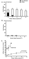
Adhesion in shear flow of K562 transfectants expressing wild-type I domains with type I or II TM domains. K562 transfectants expressing I domains with type I (C-terminal) or type II (N-terminal) TM domains were infused into the flow chamber in HBSS medium containing 1 mM Mg(II) and 1 mM Ca(II) with 10 μg/mL mouse IgG1 (control). Cells were allowed to accumulate at a wall shear stress of 0.3 dyn/cm2 for 30 s in a parallel wall flow chamber with an ICAM-1-Fcγ substrate. The wall shear stress was increased to 0.4 dyn/cm2 and incremented every 10 s. (A and B) Number of firmly adherent and rolling cells with type I (A) and type II (B) TM domains. (C) Average rolling velocity of all cells. Bars show the standard deviations of three experiments each with five replicates.
Figure 4
Figure 4
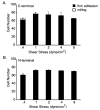
Adhesion in shear flow of K562 transfectants expressing high-affinity, mutant I domains with type I or II TM domains. K562 transfectants expressing high-affinity, K287C/K294C mutant I domains were infused exactly as described in the legend of Figure 3. (A and B) Number of firmly adherent and rolling cells with type I (A) and type II (B) TM domains. Bars show the standard deviations of three experiments each with five replicates.
Figure 5
Figure 5
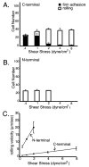
Adhesion in shear flow of microspheres decorated with N- or C-terminally linked I domains. Streptavidin-coated micro-spheres with I domains linked through C-terminal (A) or N-terminal (B) biotin tags were infused into a parallel wall flow chamber with an ICAM-1-Fcγ substrate in PBS containing 2 mM Mg(II) with 0.1% BSA and 10 μg/mL mouse IgG1 (control). Beads were allowed to accumulate at a wall shear stress of 0.3 dyn/cm2 for 30 s. The wall shear stress was increased to 0.4 dyn/cm2 and incremented every 10 s. (C) Average rolling velocity of all beads. Bars show the standard deviations of three experiments each with five replicates.
Figure 6
Figure 6
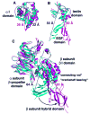
Mechanochemical design of adhesion molecules: (A) αL I domain, (B) P-selectin, and (C) αVβ3 and αIIbβ3 headpieces. High-affinity, liganded (magenta) and low-affinity, unliganded (cyan) conformations are shown superimposed using backbone regions that move little between the two conformations. Dashed lines connect a Mg2+ (A and C) or Ca2+ (B) ion shown as a sphere in the center of the ligand-binding site to the most C-terminal residue shared between the pairs of structures, which in an intact adhesion molecule would connect to other domains that tether the adhesion molecule to the cell surface. The distances between the metal in the ligand binding site and this C-terminal residue, which tensile force would tend to increase, are shown. The dashed lines and distances are red for high-affinity, liganded conformations and blue for low-affinity, unliganded conformations. Solid cylinders emphasize the axes of the α 7 helices of the α I domain (A) and β I domain (C) in each conformation. αL I domain models (A) are from ref and are based on refs and . P-Selectin lectin–EGF domain structures (B) are from ref . αVβ3 (low-affinity, unliganded) and αIIbβ3 (high-affinity, liganded) headpiece structures (C) are from refs and .

References

    1. Chen S, Springer TA. An automatic braking system that stabilizes leukocyte rolling by an increase in selectin bond number with shear. J Cell Biol. 1999;144:185–200. - PMC - PubMed
    1. Zhu C, Long M, Chesla SE, Bongrand P. Measuring receptor/ligand interaction at the single-bond level: Experimental and interpretative issues. Ann Biomed Eng. 2002;30:305–314. - PubMed
    1. Yago T, Leppanen A, Qiu H, Marcus WD, Nollert MU, Zhu C, Cummings RD, McEver RP. Distinct molecular and cellular contributions to stabilizing selectin-mediated rolling under flow. J Cell Biol. 2002;158:787–799. - PMC - PubMed
    1. Evans E, Leung A, Heinrich V, Zhu C. Mechanical switching and coupling between two dissociation pathways in a P-selectin adhesion bond. Proc Natl Acad Sci USA. 2004;101:11281–11286. - PMC - PubMed
    1. Konstantopoulos K, Hanley WD, Wirtz D. Receptor-ligand binding: ‘Catch’ bonds finally caught. Curr Biol. 2003;13:R611–R613. - PubMed

Publication types

MeSH terms