. 2007 Feb;120(2):273-80.
doi: 10.1111/j.1365-2567.2006.02498.x. Epub 2006 Nov 29.

A comparison of the binding of secretory component to immunoglobulin A (IgA) in human colostral S-IgA1 and S-IgA2

Affiliations

A comparison of the binding of secretory component to immunoglobulin A (IgA) in human colostral S-IgA1 and S-IgA2

Adel Almogren et al. Immunology. 2007 Feb.

Abstract

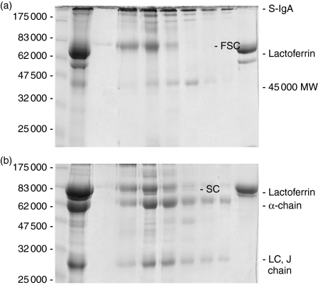
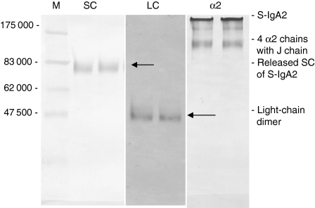
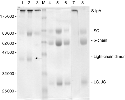
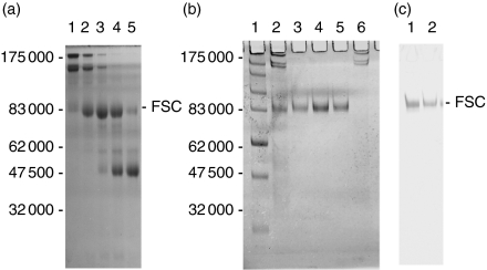
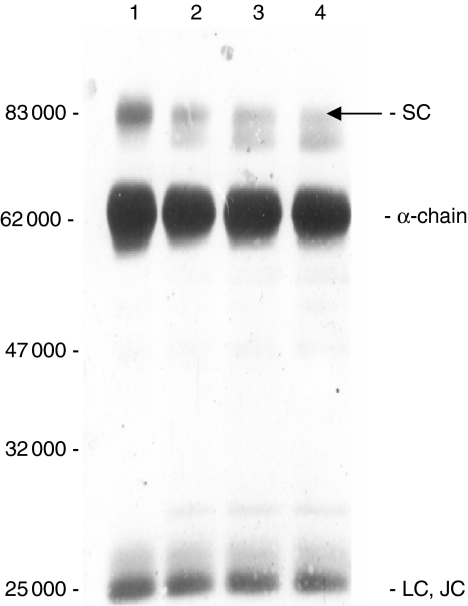
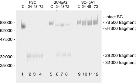
A detailed investigation of the binding of secretory component to immunoglobulin A (IgA) in human secretory IgA2 (S-IgA2) was made possible by the development of a new method of purifying S-IgA1, S-IgA2 and free secretory component from human colostrum using thiophilic gel chromatography and chromatography on Jacalin-agarose. Sodium dodecyl sulphate-polyacrylamide gel electrophoresis of unreduced pure S-IgA2 revealed that, unlike in S-IgA1, a significant proportion of the secretory component was bound non-covalently in S-IgA2. When S-IgA1 was incubated with a protease purified from Proteus mirabilis the secretory component, but not the alpha-chain, was cleaved. This is in contrast to serum IgA1, in which the alpha-chain was cleaved under the same conditions - direct evidence that secretory component does protect the alpha-chain from proteolytic cleavage in S-IgA. Comparisons between the products of cleavage with P. mirabilis protease of free secretory component and bound secretory component in S-IgA1 and S-IgA2 also indicated that, contrary to the general assumption, the binding of secretory component to IgA is different in S-IgA2 from that in S-IgA1.


Figures

Figure 1
Figure 1
Coomassie-Blue-stained 10% SDS–PAGE of clarified human colostrum and fractions from chromatography of colostrum on thiophilic resin Gels were run under (a) non-reducing conditions or (b) reducing conditions. Lane 1, protein markers; lane 2 clarified colostrum starting material; lanes 3–9, every second fraction eluted from thiophilic resin; lane 10 column run-through.
Figure 3
Figure 3
Western blot (10% SDS–PAGE run under non-reducing conditions) of dimeric S-IgA2 peak fractions from Sephacryl S-300 column probed with rabbit anti-SC antibody (SC), goat anti-human-light chains (λ and κ) antibody (LC) or 5% SDS–PAGE blotted with monoclonal anti-α2 (α2) Protein markers are shown in lane 1.
Figure 2
Figure 2
Coomassie-Blue-stained, 10% SDS–PAGE of fractions of purified S-IgA2 (lanes 1–3, 4–6) and S-IgA1 (lanes 7, 8) run under non-reducing (lanes 1–3, 7) or reducing (lanes 4–6, 8) conditions. Lane M, protein markers. The 45 000 MW protein band marked by the arrow (lanes 1, 2) is a light-chain dimer.
Figure 4
Figure 4
(a) Coomassie-Blue-stained 10% SDS–PAGE run under non-reducing conditions of fractions (lanes 1–5) from the third peak eluted from an S300 column loaded with proteins from the Jacalin–agarose column run-through. Lane 3 is the peak fraction. (b,c) FSC fractions from the S300 column pooled and chromatographed on a pIgA1-Sepharose column. Material eluted from pIgA1-Sepharose column is shown stained with Coomassie Blue (b) or after Western blot with rabbit anti-human SC antibody (c). (b) Lane 1 contains molecular weight markers; lane 2, material loaded onto column; lanes 3–5, material eluted from column; lane 6 is column run-through. (c) Lanes 1 and 2 are eluted fractions.
Figure 5
Figure 5
Coomassie-blue-stained 10% SDS–PAGE under reducing conditions of S-IgA1 incubated at 37° with either buffer alone for 40 hr (lane 1) or with P. mirabilis purified protease S-IgA1 for 8 hr (lane 2), 16 hr (lane 3) and 40 hr (lane 4). The 76 500 MW fragment derived from secretory component (SC) is indicated by the arrow.
Figure 6
Figure 6
Western blot of 10% SDS–PAGE run under reducing conditions showing structural features of bound and of untreated (control – C) free secretory component, SC-IgA2 and SC-IgA1 and these after cleavage with P. mirabilis protease at 37° for 24, 48 and 72 hr and probed with rabbit anti-human-SC antibody (detecting antibody is a goat anti-rabbit IgG conjugated with alkaline phosphatase). Cleavage of FSC and the SC in S-IgA2 was complete within 24 hr but that of the SC in S-IgA1 remained uncleaved after 72 hr.

Similar articles

Cited by

References

    1. Woof JM, Kerr MA, Moro I, Mestecky JR. Mucosal immunoglobulins. In: Mestecky JR, Bienenstock J, Lamm ME, Mayer L, Mcghee JR, Strober W, editors. Mucosal Immunology. 3. San Diego: Academic Press Inc.; 2005. pp. 153–81.
    1. Woof JM, Kerr MA. Function of immunoglobulin A in immunity. J Pathol. 2006;208:270–82. - PubMed
    1. Mostov KE. Transepithelial transport of immunoglobulins. Annu Rev Immunol. 1994;12:63–84. - PubMed
    1. Fallgreen-Gebauer E, Gebauer W, Bastian A, Kratzin HD, Eiffert H, Zimmermann B, Karas M, Hilschmann N. The covalent linkage of secretory component to IgA. Structure of sIgA. Biol Chem Hoppe Seyler. 1993;374:1023–8. - PubMed
    1. Chintalacharuvu KR, Tavill AS, Louis LN, Vaerman JP, Lamm ME, Kaetzel CS. Disulfide bond formation between dimeric immunoglobulin A and the polymeric immunoglobulin receptor during hepatic transcytosis. Hepatology. 1994;19:162–73. - PubMed

MeSH terms

LinkOut - more resources