. 2006 Dec 11:5:71.
doi: 10.1186/1476-4598-5-71.

Valproic acid and butyrate induce apoptosis in human cancer cells through inhibition of gene expression of Akt/protein kinase B

Affiliations

Valproic acid and butyrate induce apoptosis in human cancer cells through inhibition of gene expression of Akt/protein kinase B

Jihong Chen et al. Mol Cancer. .

Abstract

Background: In eukaryotic cells, the genomic DNA is packed with histones to form the nucleosome and chromatin structure. Reversible acetylation of the histone tails plays an important role in the control of specific gene expression. Mounting evidence has established that histone deacetylase inhibitors selectively induce cellular differentiation, growth arrest and apoptosis in variety of cancer cells, making them a promising class of anticancer drugs. However, the molecular mechanisms of the anti-cancer effects of these inhibitors have yet to be understood.

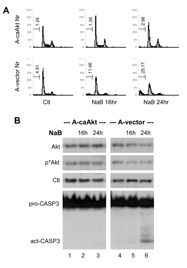
Results: Here, we report that a key determinant for the susceptibility of cancer cells to histone deacetylase inhibitors is their ability to maintain cellular Akt activity in response to the treatment. Also known as protein kinase B, Akt is an essential pro-survival factor in cell proliferation and is often deregulated during tumorigenesis. We show that histone deacetylase inhibitors, such as valproic acid and butyrate, impede Akt1 and Akt2 expression, which leads to Akt deactivation and apoptotic cell death. In addition, valproic acid and butyrate induce apoptosis through the caspase-dependent pathway. The activity of caspase-9 is robustly activated upon valproic acid or butyrate treatment. Constitutively active Akt is able to block the caspase activation and rescues cells from butyrate-induced apoptotic cell death.

Conclusion: Our study demonstrates that although the primary target of histone deacetylase inhibitors is transcription, it is the capacity of cells to maintain cellular survival networks that determines their fate of survival.


Figures

Figure 1
Figure 1
Valproic acid and butyrate repress Akt expression and induce apoptosis in HeLa cells. (A) Cell death was measured by flow cytometry analysis of subG1 population following propidium iodide staining of permibilized HeLa cells after exposure to valproic acid (VPA, 2 mM) or sodium butyrate (NaB, 5 mM) for 8, 16 or 24 hours. (B) Following 16 hours of valproic acid or butyrate treatment, the mRNA levels of Akt1, Akt2 and Akt3 in the HeLa cells were determined by quantitative real-time RT-PCR analysis with a TaqMan probe protocol. 18S rRNA was used as an internal control. Results show fold variations of treated cells in comparison to untreated controls. Error bars represent standard deviations of three independent experiments. (C) The relative mRNA abundance of Akt1, Akt2 and Akt3 isoforms of the HeLa cells was assessed by quantitative RT-PCR and plotted as fold variations of Akt1. Error bars represent the standard deviations of three independent experiments.
Figure 2
Figure 2
Deactivation of Akt by valproic acid and butyrate. (A) Equal amounts of whole cell extract (50 μg) were used to examine the levels of endogenous Akt and phospho-Akt in the HeLa cells treated with valproic acid (VPA, 2 mM) or sodium butyrate (NaB, 5 mM) for 16 or 24 hours. The blot was then stripped and reprobed with a SRC antibody for protein loading controls. (B) Quantitative analysis of the Akt blots is expressed as fold variations compared to untreated control after being normalized to the loading controls. Error bars represent standard deviations of three independent experiments. (C) The quantification of phospho-Akt Western blots was performed as described for panel B.
Figure 3
Figure 3
Valproic acid and butyrate induce caspase activation. (A) Equal amounts of whole cell extracts (50 μg) were used for Western blot analysis of caspase-3 in HeLa cells upon treatment with valproic acid (VPA, 2 mM) or sodium butyrate (NaB, 5 mM) for 16 or 24 hours. The blots were then stripped and reprobed with a β-actin antibody for protein loading controls. (B) The level of caspase-9 mRNA in the HeLa cells was examined by quantitative real-time RT-PCR analysis following valproic acid or butyrate treatment. Error bars represent standard deviations of three independent experiments. (C) and (D) Following 16 hours of valproic acid or butyrate treatment, the HeLa cells were harvested and assayed for caspase-9 and caspase-8 activities in parallel. Results show fold induction of the activities relative to untreated controls after background subtraction of zero time signals. Error bars represent the standard deviations of five independent experiments.
Figure 4
Figure 4
Constitutively active Akt counteracts butyrate-induced apoptosis. (A) Ovarian carcinoma A2780S cell lines stably integrated with an expression plasmid for constitutively active Akt (A-caAkt) or an empty vector (A-vector) were used for flow cytometry analysis of cells stained with propidium iodide after 16 or 24 hours exposure to sodium butyrate (NaB, 5 mM). The percentage of cells in the subG1 population is indicated in the corresponding graphs. Untreated cells were used as controls. (B) Equal amounts of whole cell extracts (50 μg) were used for Western blot analysis of Akt, phospho-Akt and casepase-3 levels in the A-caAkt and A-vector cells following butyrate treatments. The blot was then stripped and reprobed for loading control (Ctl) with a SRC antibody.
Figure 5
Figure 5
SiHa cell survival is not affected by valproic acid and butyrate. (A) Flow cytometry analysis of propidium iodide uptake of the SiHa cells following exposure to valproic acid (VPA, 2 mM) or sodium butyrate (NaB, 5 mM). Untreated SiHa cells were used as control and the time of treatments was for 16 or 24 hours. (B) Equal amounts of whole cell extracts (50 μg) were used for Western blot analysis of endogenous Akt and phospho-Akt of the SiHa cells following treatment with valproic acid or butyrate for 16 or 24 hours. The blot was then stripped and reprobed for loading control (Ctl) with a SRC antibody. (C) and (D) Following 16 hours of valproic acid or butyrate treatment, the mRNA levels of Akt1, Akt2 and Akt3 in the SiHa cells were examined by quantitative real-time RT-PCR analysis. 18S was used as an internal control. Results show fold variations of treated cells compared to untreated controls. Error bars represent standard deviations of three independent experiments.
Figure 6
Figure 6
The Akt mRNA levels and caspase activities of the SiHa cells. (A) The relative mRNA abundance of Akt1, Akt2 and Akt3 in the HeLa and SiHa cells were assessed by quantitative RT-PCR and plotted as fold variations of Akt1 mRNA level of the HeLa cells. 18S was used as an internal control. Error bars represent standard deviations of three independent experiments. (B) The relative level of Akt3 mRNA following 16 hours of exposure to valproic acid (VPA, 2 mM) or sodium butyrate (NaB, 5 mM) was plotted as arbitrary units (A.U.) of the mRNA abundance. (C) The level of caspase-9 mRNA in the SiHa cells was examined by quantitative real-time RT-PCR analysis following valproic acid or butyrate treatment. Results are expressed as fold induction of the transcripts relative to untreated controls. Error bars represent standard deviations of three independent experiments. (D) The experimental set up was as in panel A except that the caspase-9 mRNA level in untreated SiHa cells was compared to untreated HeLa cells. (E) Following 16 hours of valproic acid or butyrate treatment, the SiHa cells were assayed for their caspase-8 and caspase-9 activities. Results show fold induction of the activities relative to untreated controls after background subtraction of zero time signals. Error bars represent standard deviations of five independent experiments.

References

    1. Janknecht R, Hunter T. Versatile molecular glue. Transcriptional control. Curr Biol. 1996;6:951–954. doi: 10.1016/S0960-9822(02)00636-X. - DOI - PubMed
    1. Wong J, Li Q, Levi BZ, Shi YB, Wolffe AP. Structural and functional features of a specific nucleosome containing a recognition element for the thyroid hormone receptor. EMBO J. 1997;16:7130–7145. doi: 10.1093/emboj/16.23.7130. - DOI - PMC - PubMed
    1. Wolffe AP. Histones, nucleosomes and the roles of chromatin structure in transcriptional control. Biochem Soc Trans. 1997;25:354–358. - PubMed
    1. Li Q, Wrange O. Accessibility of a glucocorticoid response element in a nucleosome depends on its rotational positioning. Mol Cell Biol. 1995;15:4375–4384. - PMC - PubMed
    1. Li Q, Wrange O. Translational positioning of a nucleosomal glucocorticoid response element modulates glucocorticoid receptor affinity. Genes Dev. 1993;7:2471–2482. - PubMed

Publication types

LinkOut - more resources