. 2007 Jan 19;1129(1):63-71.
doi: 10.1016/j.brainres.2006.09.112. Epub 2006 Dec 6.

Transient electrical coupling regulates formation of neuronal networks

Affiliations

Transient electrical coupling regulates formation of neuronal networks

Theresa M Szabo et al. Brain Res. .

Abstract

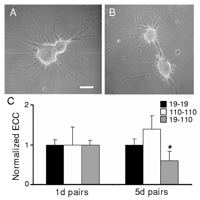
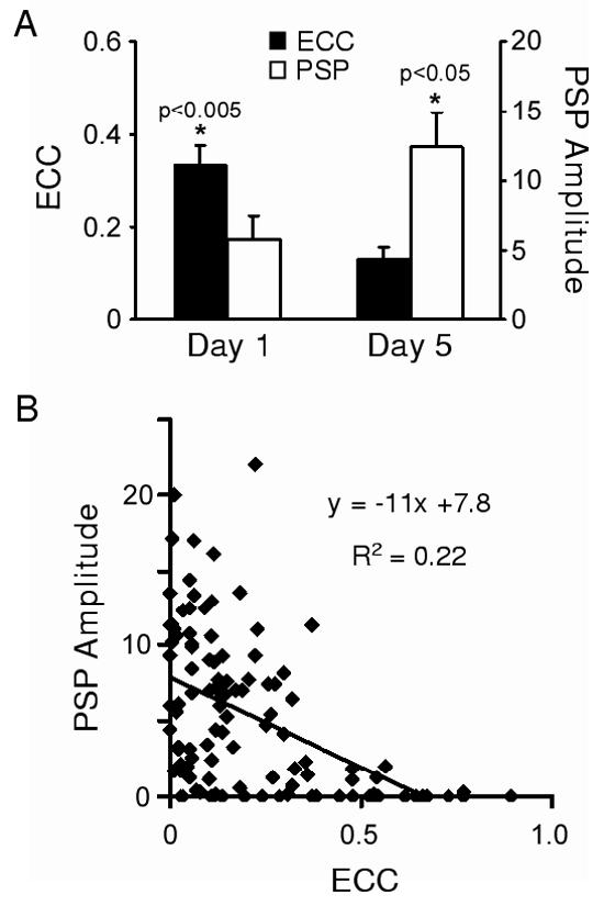
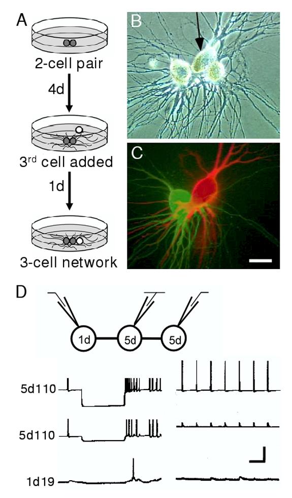
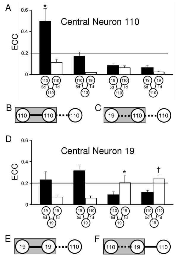
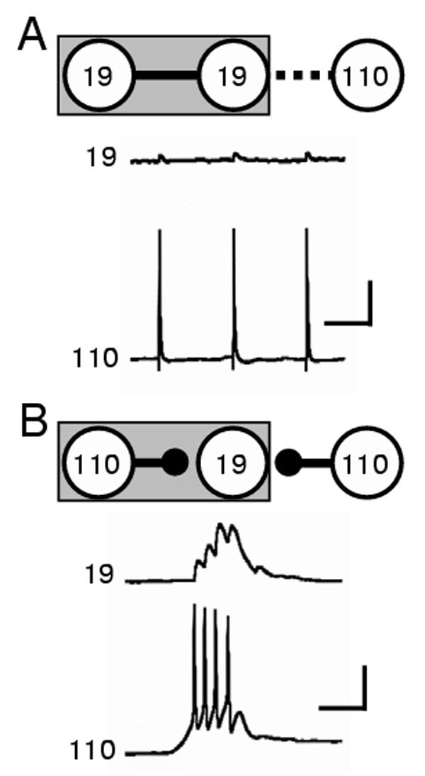
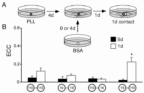
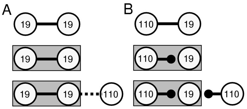
Electrical synapses are abundant before and during developmental windows of intense chemical synapse formation, and might therefore contribute to the establishment of neuronal networks. Transient electrical coupling develops and is then eliminated between regenerating Helisoma motoneurons 110 and 19 during a period of 48-72 h in vivo and in vitro following nerve injury. An inverse relationship exists between electrical coupling and chemical synaptic transmission at these synapses, such that the decline in electrical coupling is coincident with the emergence of cholinergic synaptic transmission. In this study, we have generated two- and three-cell neuronal networks to test whether predicted synaptogenic capabilities were affected by previous synaptic interactions. Electrophysiological analyses demonstrated that synapses formed in three-cell neuronal networks were not those predicted based on synaptogenic outcomes in two-cell networks. Thus, new electrical and chemical synapse formation within a neuronal network is dependent on existing connectivity of that network. In addition, new contacts formed with established networks have little impact on these existing connections. These results suggest that network-dependent mechanisms, particularly those mediated by gap junctional coupling, regulate synapse formation within simple neural networks.


Figures

Figure 1
Figure 1
Formation of electrical synaptic connections between neurons paired in outgrowth-permissive conditions. (A) Neurons were plated in contacting pairs for 5d on PLL-coated dishes, which promoted adherence to the dish surface and outgrowth of processes. After 5d of outgrowth, neurons 19 (larger soma) and 110 exhibited extensive neuritic processes. (B) Although neurons were plated with contacting somata, in some pairs somata moved apart as a large fascicle of processes developed between the two cell bodies. (C) Electrical coupling coefficients (ECCs) were determined as the ratio of postsynaptic to presynaptic voltage changes. ECC values (normalized mean ± sem) for 19-19, 110-110, and 19-110 pairs, with 5d ECC values normalized to 1d values. (19-19: 1d ECC = 0.32 ± 0.06, n = 29 vs. 5d ECC = 0.32 ± 0.07, n = 28; NSD (no significant difference). 110-110: 1d ECC = 0.11 ± 0.05, n = 6 vs. 5d ECC = 0.15 ± 0.05, n = 6; NSD. 19-110: 1d ECC = 0.33 ± 0.06, n = 32 vs. 5d ECC = 0.20 ± 0.05, n = 19; *, p<0.05, student’s t-test).
Figure 2
Figure 2
Temporal expression of chemical and electrical synaptic connections. (A) Histogram illustrating electrical coupling coefficients (ECC) and PSP amplitude measured at 95 msec after the peak of the presynaptic action potential on day 1 (n=32) and day 5 (n=19) in culture. Both the reduction in ECC values and increase in PSP amplitudes were significant (ECC: p<0.005; PSP: p<0.05; one-tailed student’s t-tests). (B) Scatterplot of 19-110 pairs from (A) and a linear fit of the relationship between ECC and PSP amplitude (n=51).
Figure 3
Figure 3
Experimental paradigm for investigating three-cell networks in culture. (A) Cells were plated into adhesive cell culture dishes in pairs (grey spheres). Following 4 days of contact and neurite outgrowth a third, younger cell (white sphere) was plated into contact with one cell of the pair for an additional day. (B) Phase-contrast micrograph showing neuronal somata plated in a row, with one central cell (arrow) paired to an older cell on one side, and a younger cell on the other. (C) Fluorescent dye-fill of the three-cell network in A. The central neuron 19 has been injected with Texas Red and the left neuron 110 with Lucifer yellow. Scale bar equals 30 μm. (D) Schematic of the recording configuration (top) illustrates that current was injected in one neuron of a network while simultaneous recordings from the other two cells were used to assess network connectivity. Hyperpolarizing (lower left traces) and depolarizing (lower right traces) current injections into one neuron (top traces) permitted assessment of connectivity with its 1 and 5d neighbors. In this case, hyperpolarizing current injected into a central 5d 110 elicited a hyperpolarization in a 5d 110 neighbor, but only weak electrical coupling was detected in the 1d 19 partner. Depolarizing current injection demonstrated weak chemical connectivity between the 5d 110 and the 1d 19. Note that the events recorded in the 5d 110 (middle trace, right panel) represent electrically-mediated synaptic potentials. Chemically-mediated synapses are actually inhibitory, but appear as depolarizing potentials due to the use of KCl electrodes that alter the chemical driving forces underlying these acetylcholine-gated channels (see Szabo et al., 2004). Vertical bar equals 40 mV (left traces), 20 mV (right traces). Horizontal bar equals 1 sec.
Figure 4
Figure 4
Electrical coupling between neurons in three-cell networks. (A) Histogram of ECC values for 5d (solid bars) and 1d connections (open bars) for three-cell networks with 110 as the central neuron (from left to right, 110-110-110, n=6; 110-110-19, n=9; 19-110-110, n=7; 19-110-110, n=17). (B and E) When electrical coupling was strong (solid line) at homotypic 5d contacts, newly formed 1d connections with a neuron 110 were weak (dashed line). Grey rectangle indicates 5d neuronal contacts. (C) When neuron 110 was the central cell of a heterotypic pair, it exhibited weak coupling at both its 5d and 1d contacts. (D) Histogram of ECC values for 5d and 1d connections with 19 as the central neuron (19-19-19, n=5; 19-19-110, n=8; 110-19-5d19, n=8; 110-19-110, n=14). An analysis across all 5d homotypic contacts yielded no significant differences, except at 110-110 connections (ANOVA, *, p<0.05). An analysis across all 5d heterotypic groups yielded no significant differences, (ANOVA, p=0.48). An analysis across all 1d contact groups indicated a significant difference (ANOVA, p<0.0001) at 5d19-1d19 (post hoc LSD,*, p<0.05) and 5d19-1d110 (post hoc LSD, †, p<0.05). (F) Heterotypic 5d contacts, where neuron 19 was the central cell, exhibited weak coupling and formed strong electrical connections at new 110 contacts. Overall, the presence of strong electrical coupling (e.g., B and E) appears to limit formation of new electrical synapses, while the presence of weak coupling (e.g., C and F) affected subsequent electrical synaptogenesis in a cell-specific manner.
Figure 5
Figure 5
Chemical synapse formation between specific neurons is network dependent. (A) Schematic and sample traces illustrating one example of synaptogenesis in a 3-cell network: a 5d 19-19 contact possessed strong electrical coupling (solid line in schematic) and no chemical synaptic transmission, while only weak electrical coupling (dashed line) and no chemical synaptic transmission was recorded at the 1d contact of the central 19 and 110 (record traces). Scale bars equal 20mV and 1 sec. (B) By 5 days in culture, the strength of electrical connections between central neuron 19 and neuron 110 had diminished and strong chemical synaptic transmission was present (solid circle head). Strong chemical connections were also formed at the 1d contact between neurons 19 and 110 in this three-cell configuration (record traces). Scale bars equal 20mV and 1 sec.
Figure 6
Figure 6
Effects of cell age on electrical synapse formation. (A) Schematic of the experimental paradigm. Cells were plated onto PLL-coated dishes and allowed to extend processes (dark spheres). On d4, cells were contacted by new somata (white sphere) that had been cultured in separate non-adhesive conditions for either 0 or 4 days. After 24 hours of contact, synaptic connectivity was assessed. (B) Histogram illustrating ECC values for “equally-aged” contacts (5d, solid bars) and “differently-aged” contacts (1d, open bars). 5d pairs were not significantly different (ANOVA, p=0.8; from left to right, 110-110, n=7; 19-19, n=6; 110-19, n=9; 19-110, n=8.) The 1d contact between a 5d 19 and a 1d 110 was significantly different from all other groups (ANOVA (post-hoc LSD), *, p<0.002; 110-110, n=8; 19-19, n=8; 110-19, n=10; 19-110, n=10).
Figure 7
Figure 7
Summary of key observations of chemical synapse formation in three-cell networks. (A) Electrical coupling was strong (solid line) at homotypic 19-19 contacts following 24h of contact (top diagram). At day 4 (middle diagram), strong electrical coupling was maintained. At newly formed 1d connections with a neuron 110 (bottom diagram), only weak electrical coupling (dashed line) was observed between neurons 19 and 110. (B) Heterotypic 1d contacts possessed strong electrical coupling (top diagram). However, by day 4 uncoupling had occurred and strong chemical connections were present (middle diagram). At newly formed 1d connections on day 5 (bottom diagram), neuron 110 formed strong chemical connections with central neuron 19. Note that the new neuron 110 is forming very different kinds of synaptic connections, strong chemical in B and weak electrical in A, with the same postsynaptic neuron 19. The only difference in the postsynaptic neuron is its other existing network connectivity.

References

    1. Allen F, Warner A. Gap junctional communication during neuromuscular junction formation. Neuron. 1991;6:101–111. - PubMed
    1. Arumugam H, Liu X, Colombo PJ, Corriveau RA, Belousov AB. NMDA receptors regulate developmental gap junction uncoupling via CREB signaling. Nat Neurosci. 2005;8:1720–1726. - PubMed
    1. Bem T, Le Feuvre Y, Simmers J, Meyrand P. Electrical coupling can prevent expression of adult-like properties in an embryonic neural circuit. J Neurophysiol. 2002;87:538–47. - PubMed
    1. Bennett MVL. Electrical transmission: a functional analysis and comparison to chemical transmission. In: Kandel ER, editor. Handbook of physiology. Amer Phys Society; Bethesda, MD: 1977. pp. 357–416.
    1. Bennett MVL, Zukin RS. Electrical coupling and neuronal synchronization in the mammalian brain. Neuron. 2004;41:495–511. - PubMed

Publication types

MeSH terms