

Review
doi: 10.1016/j.semcdb.2006.10.001.
Epub 2006 Oct 27.
Signaling during lens regeneration
Affiliations
- PMID: 17157042
- PMCID: PMC1828075
- DOI: 10.1016/j.semcdb.2006.10.001
Item in Clipboard
Review
Signaling during lens regeneration
Semin Cell Dev Biol.
2006 Dec.
Abstract
The newt is one of the few organisms that is able to undergo lens regeneration as an adult. This review will examine the signaling pathways that are involved in this amazing phenomenon. In addition to outlining the current research involved in elucidating the key signaling molecules in lens regeneration, we will also highlight some of the similarities and differences between lens regeneration and development.
Figures
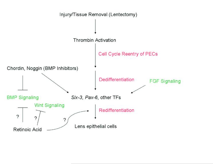
Outline of events and pathways likely to be involved in lens regeneration. Upon lens removal or injury, PECs must reenter the cell cycle and thrombin is suspected to play a role in this. Following proliferation, the PECs dedifferentiate and then redifferentiate. The BMP, Wnt, and FGF pathways play pivotal roles in lens development. BMP antagonists have been shown to upregulate TFs such as six3 in the newt during regeneration as well as induce lens regeneration. RA has been shown to inhibit BMP and Wnt signaling in other systems, but the mechanism through which it works in inducing (along with six3) newt lens regeneration is unclear. Question marks depict possible roles of RA for induction of lens regeneration.
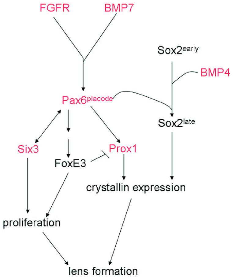
A diagram showing several factors involved in lens development. Some of these factors have also been examined in lens regeneration and are depicted with red color in the pathway. This might suggest conservation of function even though the inductive signals might be different.
Similar articles
-
Lens regeneration from the cornea requires suppression of Wnt/β-catenin signaling.Exp Eye Res. 2016 Apr;145:206-215. doi: 10.1016/j.exer.2016.01.003. Epub 2016 Jan 8. Exp Eye Res. 2016. PMID: 26778749 Free PMC article.
-
Cell signaling pathways in vertebrate lens regeneration.Curr Top Microbiol Immunol. 2013;367:75-98. doi: 10.1007/82_2012_289. Curr Top Microbiol Immunol. 2013. PMID: 23224710 Free PMC article. Review.
-
A critical role for thrombin in vertebrate lens regeneration.Philos Trans R Soc Lond B Biol Sci. 2004 May 29;359(1445):765-76. doi: 10.1098/rstb.2004.1467. Philos Trans R Soc Lond B Biol Sci. 2004. PMID: 15293804 Free PMC article. Review.
-
Eye on regeneration.Anat Rec B New Anat. 2005 Nov;287(1):42-8. doi: 10.1002/ar.b.20084. Anat Rec B New Anat. 2005. PMID: 16308862 Free PMC article. Review.
-
Unique expression patterns of the retinoblastoma (Rb) gene in intact and lens regeneration-undergoing newt eyes.Anat Rec A Discov Mol Cell Evol Biol. 2003 Mar;271(1):185-8. doi: 10.1002/ar.a.10023. Anat Rec A Discov Mol Cell Evol Biol. 2003. PMID: 12552633
Cited by
-
Molecular and cellular aspects of amphibian lens regeneration.Prog Retin Eye Res. 2010 Nov;29(6):543-55. doi: 10.1016/j.preteyeres.2010.07.002. Epub 2010 Jul 16. Prog Retin Eye Res. 2010. PMID: 20638484 Free PMC article.
-
Dedifferentiated follicular granulosa cells derived from pig ovary can transdifferentiate into osteoblasts.Biochem J. 2012 Oct 15;447(2):239-48. doi: 10.1042/BJ20120172. Biochem J. 2012. PMID: 22839299 Free PMC article.
-
Lens regeneration from the cornea requires suppression of Wnt/β-catenin signaling.Exp Eye Res. 2016 Apr;145:206-215. doi: 10.1016/j.exer.2016.01.003. Epub 2016 Jan 8. Exp Eye Res. 2016. PMID: 26778749 Free PMC article.
-
A focus on the optical properties of the regenerated newt lens.PLoS One. 2013 Aug 22;8(8):e70845. doi: 10.1371/journal.pone.0070845. eCollection 2013. PLoS One. 2013. PMID: 23990914 Free PMC article.
-
Insights into Bone Morphogenetic Protein-(BMP-) Signaling in Ocular Lens Biology and Pathology.Cells. 2021 Sep 30;10(10):2604. doi: 10.3390/cells10102604. Cells. 2021. PMID: 34685584 Free PMC article. Review.
References
-
- Eguchi G. Electron microscopic studies on lens regeneration: I, mechanisms of depigmentatioon of the iris. Embryologia. 1963;8:45–62.
-
- Tsonis PA. Regeneration in vertebrates. Dev Biol. 2000;221:273–284. - PubMed
-
- Eguchi G. Electron microscopic studies on lens regeneration: II, formation and growth of lens vesicle and differentiation of lens fibers. Embryologia. 1964;8:247–287.
-
- Yamada T. Control mechanisms in cell-type conversion in newt lens regeneration. Krager; Basel: 1977. - PubMed
-
- Walsh PN, Schmaier AH. Platelet-coagulant protein interactions. In: Colman RW, Hirsh J, Marder VJ, Salzman EW, editors. Hemostasis and Thrombosis: Basic Principles and Clinical Practice. 3rd ed. J.B. Lippincott Co.; Philadelphia: 1994. pp. 629–651.
Publication types
MeSH terms
Grants and funding
LinkOut - more resources
Full Text Sources
