. 2007 Jan 1;42(1):32-43.
doi: 10.1016/j.freeradbiomed.2006.09.014. Epub 2006 Sep 19.

Mitochondrial DNA deletions inhibit proteasomal activity and stimulate an autophagic transcript

Affiliations

Mitochondrial DNA deletions inhibit proteasomal activity and stimulate an autophagic transcript

Mansour Alemi et al. Free Radic Biol Med. .

Abstract

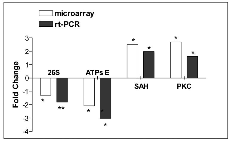
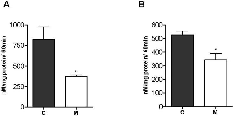
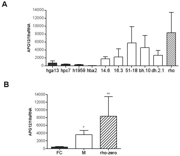
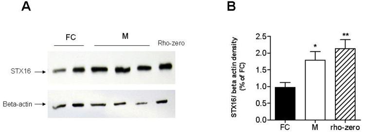
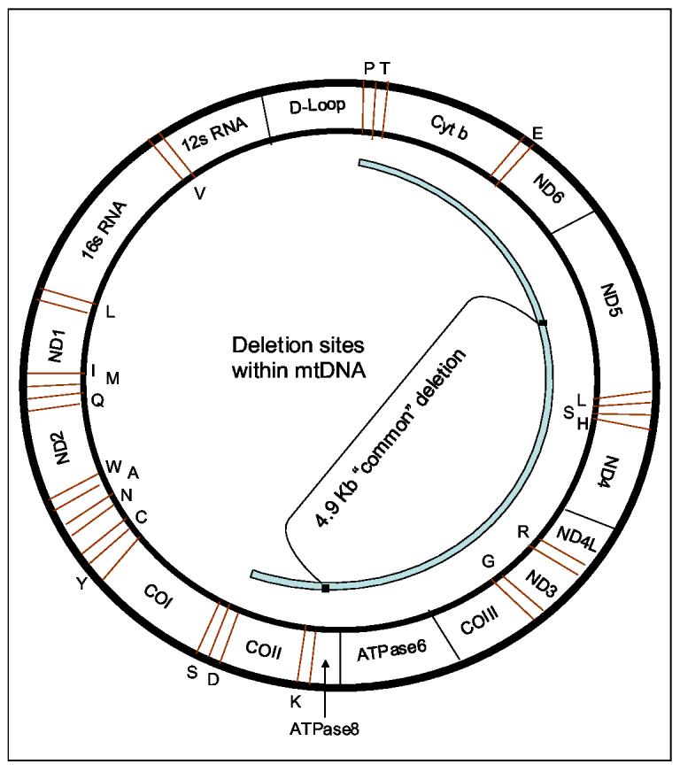
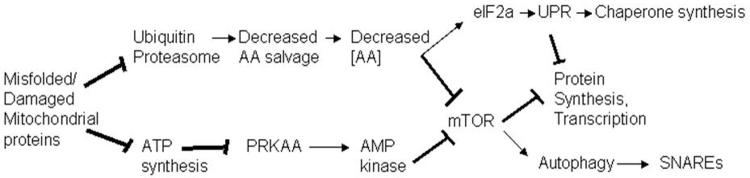
Deletions within the mitochondrial DNA (mtDNA) cause Kearns Sayre syndrome (KSS) and chronic progressive external opthalmoplegia (CPEO). The clinical signs of KSS include muscle weakness, heart block, pigmentary retinopathy, ataxia, deafness, short stature, and dementia. The identical deletions occur and rise exponentially as humans age, particularly in substantia nigra. Deletions at >30% concentration cause deficits in basic bioenergetic parameters, including membrane potential and ATP synthesis, but it is poorly understood how these alterations cause the pathologies observed in patients. To better understand the consequences of mtDNA deletions, we microarrayed six cell types containing mtDNA deletions from KSS and CPEO patients. There was a prominent inhibition of transcripts encoding ubiquitin-mediated proteasome activity, and a prominent induction of transcripts involved in the AMP kinase pathway, macroautophagy, and amino acid degradation. In mutant cells, we confirmed a decrease in proteasome biochemical activity, significantly lower concentration of several amino acids, and induction of an autophagic transcript. An interpretation consistent with the data is that mtDNA deletions increase protein damage, inhibit the ubiquitin-proteasome system, decrease amino acid salvage, and activate autophagy. This provides a novel pathophysiological mechanism for these diseases, and suggests potential therapeutic strategies.


Figures

Figure 1
Figure 1
Quantitative rt-PCR validation of microarray data. Fold changes of 4 selected genes obtained by microarray analysis and by quantitative rt-PCR are shown. QRT-PCR results were normalized to beta-actin expression. Significant differences from the control groups: * unpaired t test, p<0.05, ** unpaired t test, p<0.005.
Figure 2
Figure 2
Measurement of proteasome activity in 143B cybrids and lymphoblasts. In vitro assessment of 20S proteasome activity was done by measuring the hydrolysis of the fluorogenic peptidyl substrate Suc-Leu-Leu-Val-Tyr-AMC by the SDS-activated proteasome. (A) Average activity in three 143B fusion controls and two osteosarcoma cybrids 3.6 and 16.3 with 70% of 7,5 kb mtDNA deletions. (B) Average activity in lymphoblast cell lines from three control subjects, and two patient cell lines, (848) with 57% of 4,9 kb mtDNA deletion and (63358) with 27% of 7,4 kb mtDNA deletion. C, control cells; M, mutant cells. Error bars represent SEM. * Unpaired t test, p<0.02.
Figure 3
Figure 3
ATG12 RNA overexpression in mutant 143B cybrids Quantitative rt-PCR analysis of ATG12 RNA expression in 143B osteosarcoma cybrids. QRT-PCR results were normalized to 18sRNA expression. (A) QRT-PCR results of three separate experiments each done in duplicate. Black bars represent fusion control cell lines (FC), white bars mutant cell lines (M) and striped bars mtDNA depleted rho-zero 143B cell lines, cell line mutation status is described in Table 7. (B) Average relative ATG12 RNA expression of fusion controls, mutant cybrids and 143B rho-zero cells at left, summed. Error bars represent SEM. * Unpaired t test, p<0.05. ** Unpaired t test, p<0.01.
Figure 4
Figure 4
Syntaxin 16 protein overexpression in mutant 143B cybrids. Relative expression of syntaxin 16 (STX16) in 143B mutant cybrids (M) compared to 143B fusion control cybrids (FC). (A) Representative immunoblot for STX16 out of three separate experiments. From left, the samples were: FC (fusion controls)-HGA13 & HPC7; M-Mutants: 14.6, 16.3. and 51-18. The blots were stripped and reprobed against beta-actin. (B) Average relative density (syntaxin 16 over beta-actin) in fusion controls, mutant cybrids and 143B rho-zero cells. Error bars represent SEM. * Unpaired t test, p<0.05. ** Unpaired t test, p<0.005.
Figure 5
Figure 5
Deletion sites within mtDNA. Deletions of mtDNA may occur within a certain region (shown in light blue). The deleted genes code for respiratory chain subunits of Complex I (ND3, ND4, ND4L, ND5 and ND6), Complex III (Cyt b), Complex IV (COII and COIII), and Complex V (ATPase 6and 8). The deletions may erase also transfer RNA genes (indicated in red) such as S, D, K, G, R, H, S, L, E, T, and P.
Figure 6
Figure 6
A mechanistic hypothesis for the consequences of mtDNA deletions. Expected consequences of mtDNA deletions. Deletions must perforce erase essential tRNAs and components of multisubunit complexes of the OXPHOS chain, and result in increased protein oxidative damage. Increased protein oxidative damage has been demonstrated to inhibit ubiquitin-proteasomal activity, which is expected to decrease the rate of salvage of amino acids, and result in decreased amino acid concentration in cells, which has previously been demonstrated to decrease the activity of mTOR, which then represses protein synthesis and activates autophagy. Deletions were shown to result in decreased expression of nuclear-encoded OXPHOS complex (of both the electron transport chain and ATP synthase), which must also inhibit ATP synthesis (which has been demonstrated at the biochemical level by others), and stimulate the activity of AMP kinase, which then should inhibit mTOR, which should result in the inhibition of protein synthesis and the activation of autophagy [52].

Comment in

References

    1. Holt IJ, Harding AE, Morgan-Hughes JA. Deletions of muscle mitochondrial DNA in patients with mitochondrial myopathies. Nature. 1988;331:717–719. - PubMed
    1. Moraes CT, DiMauro S, Zeviani M, Lombes A, Shanske S, Miranda AF, Nakase H, Bonilla E, Werneck LC, Servidei S, et al. Mitochondrial DNA deletions in progressive external ophthalmoplegia and Kearns-Sayre syndrome. N. Engl. J. Med. 1989;320:1293–1299. - PubMed
    1. Rotig A, Cormier V, Blanche S, Bonnefont JP, Ledeist F, Romero N, Schmitz J, Rustin P, Fischer A, Saudubray JM, et al. Pearson's marrow-pancreas syndrome. A multisystem mitochondrial disorder in infancy. J. Clin. Invest. 1990;86:1601–1608. - PMC - PubMed
    1. Johns DR, Rutledge SL, Stine OC, Hurko O. Directly repeated sequences associated with pathogenic mitochondrial DNA deletions. Proc. Natl. Acad. Sci. U S A. 1989;86:8059–8062. - PMC - PubMed
    1. Schon EA, Rizzuto R, Moraes CT, Nakase H, Zeviani M, DiMauro S. A direct repeat is a hotspot for large-scale deletion of human mitochondrial DNA. Science. 1989;244:346–349. - PubMed

Publication types

MeSH terms