. 2006 Dec 8;24(5):785-796.
doi: 10.1016/j.molcel.2006.10.026.

Yng1 PHD finger binding to H3 trimethylated at K4 promotes NuA3 HAT activity at K14 of H3 and transcription at a subset of targeted ORFs

Affiliations

Yng1 PHD finger binding to H3 trimethylated at K4 promotes NuA3 HAT activity at K14 of H3 and transcription at a subset of targeted ORFs

Sean D Taverna et al. Mol Cell. .

Abstract

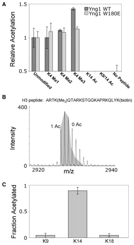
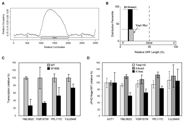
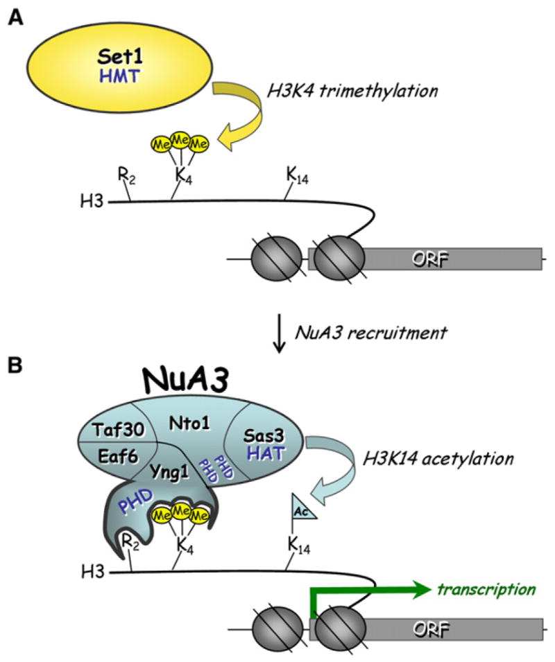
Posttranslational histone modifications participate in modulating the structure and function of chromatin. Promoters of transcribed genes are enriched with K4 trimethylation and hyperacetylation on the N-terminal tail of histone H3. Recently, PHD finger proteins, like Yng1 in the NuA3 HAT complex, were shown to interact with H3K4me3, indicating a biochemical link between K4 methylation and hyperacetylation. By using a combination of mass spectrometry, biochemistry, and NMR, we detail the Yng1 PHD-H3K4me3 interaction and the importance of NuA3-dependent acetylation at K14. Furthermore, genome-wide ChIP-Chip analysis demonstrates colocalization of Yng1 and H3K4me3 in vivo. Disrupting the K4me3 binding of Yng1 altered K14ac and transcription at certain genes, thereby demonstrating direct in vivo evidence of sequential trimethyl binding, acetyltransferase activity, and gene regulation by NuA3. Our data support a general mechanism of transcriptional control through which histone acetylation upstream of gene activation is promoted partially through availability of H3K4me3, "read" by binding modules in select subunits.


Figures

Figure 1
Figure 1. Isolation of an Yng1-Containing NuA3 Protein Complex
(A) Isolation and identification of an Yng1-TAP-containing NuA3 protein complex. IgG-coated Dynabeads were incubated with S. cerevisiae lysate from either a strain containing no affinity tag or a strain containing TAP-tagged Yng1. The Yng1-TAP lysate contained an equal amount of d4-lysine-labeled cell lysate (untagged) for I-DIRT analysis. Yng1-TAP and associating proteins were resolved by 4%–12% denaturing gel electrophoresis, visualized by Coomassie staining, and excised for mass spectrometric protein identification. Proteins associated with an in-genome mutated version of Yng1 (W180E) were also identified (see text for details). (B) I-DIRT analysis of the Yng1-TAP-associated proteins identified in (A). Proteins identified as containing near 100% h4-lysine (isotopically light) are true protein complex components, while protein identifications containing 50% h4-lysine (and therefore 50% d4-lysine) are contaminants. Error bars show the standard deviation for lysine-containing peptides. (C) Schematic representation of proteins identified in (B) as stable components of the Yng1-TAP-containing protein complex.
Figure 2
Figure 2. NuA3 Is Targeted to H3 K4me3 by Yng1
(A) Peptide pull-down assays were performed by using yeast lysates from tagged strains and biotinylated H3 peptides (either unmodified, monomethylated, dimethylated, or trimethylated at K4) bound to streptavidin-linked Dynabeads. In all cases, pull-downs were analyzed with antibodies recognizing the PrA epitope in the TAP tag. (B) NuA3 binding to H3K4me3 is context specific. Lysates from wild-type or Yng1 W180E strains were processed as in (A), and peptide pull-downs were performed with the H3K4me3peptides indicated. (C) The binding of NuA3 to H3K4me3 peptide is directed solely through the PHD finger of Yng1. Lysates from knockout strains indicated were processed and pull-downs were performed as in (A). The asterisk on (Cc) represents a breakdown product of the tagged Yng1.
Figure 3
Figure 3. Yng1 Binds Directly to H3 K4me3 through Its Single PHD Finger
(A) Peptide pull-down assays were performed using cryogenically processed yeast, and the pull-down lane was compared to the input lane to gauge enrichment. As expected, anti-GST antibodies show that GST fusions were enriched in glutathione pull-downs (see Ag). (B) A titration plot based on the chemical shift changes of the W180 side chain imino resonance from PHD domain of YNG1 protein. Chemical shift changes were measured by 1H15N-HSQC spectra upon addition of monomethylated, dimethylated, trimethylated, and unmodified lysine 4 from histone H3 peptide as a function of the molar ratio. The concentrated peptide (20 mM) was added at the molar ratio of 0.5, 1.0, 1.5, 2.2, 3.0, and 5.0 peptide to protein. Δδ was calculated based on the method described in Supplemental Experimental Procedures. (C) Fluorescence anisotropy was used to determine the dissociation constants for the interaction between the Yng1 PHD finger and different methylation states of H3K4. Error bars are the standard deviation from triplicate analyses. (D) Tabulated values for the dissociation constants measured in Figure 3C.
Figure 4
Figure 4. NMR Structure of the Yng1 PHD Interaction with Trimethylated H3 K4
NMR-derived structure of the PHD finger (152–212) in the free form. For clarity, residues 152–154 and 208–212 in the unstructured regions were omitted. (A) Backbone superposition of 20 energy-minimized structures of the PHD finger. (B) Aromatically rich surface that shows extensive chemical shift changes upon peptide binding (Y157 and W180). (C) Backbone superposition of 20 energy-minimized structures of the PHD finger in complex with H31–9K4me3 peptide. (D) Aromatically rich surface that shows extensive interactions with trimethylated lysine K4 (Y157 and W180). (E) Surface representation of the YNG1 PHD complex with H31–9K4me3 peptide. Surface residues that undergo the largest chemical shift upon binding are highlighted in pink. (F) An ensemble of 20 structures with side chains involved in complex formation colored in purple (D172 and E179 for H3 R2, Y157 and W180 for H3K4).
Figure 5
Figure 5. HAT Activity of the Yng1-TAP-Containing Protein Complex
(A) NuA3 exhibits enhanced HAT activity upon trimethylation of H3K4. Peptide-eluted Yng1-TAP-containing protein complex was incubated with differentially modified versions of histone H31–20 peptide and with radiolabeled acetyl CoA. Data were normalized to the amount of acetylation observed for the unmodified peptide. (B) Yng1-TAP-containing NuA3 protein complex incorporated one acetyl group per peptide. Peptide-eluted Yng1-TAP containing NuA3 protein complex was incubated with H3K4me3 peptide and acetyl CoA. Following the HAT reaction, unacetylated lysines were chemically acetylated with d6-acetic anhydride. The mass spectrum from this reaction showed that only one acetylation was detectable on any given peptide. The shaded area shows the theoretical isotopic distribution from the singly acetylated H3 peptide. Peak area excluded from the shaded area is due to the unacetylated version of the H3 peptide. (C) The peptide-eluted Yng1-TAP-containing NuA3 protein complex preferentially acetylated the input H3K4me3 peptide on K14. Shown is a plot of the fraction of acetylation at each modifiable lysine on the peptide. The fraction acetylated was determined by mass spectrometric fragmentation of the singly acetylated peptide from (B). Error bars are the standard deviation from triplicate analyses.
Figure 6
Figure 6. Genome-Wide Localization of Yng1-Myc, Its Transcriptional Impact, and Its Epigenetic Effect
(A) YNG1 was genomically MYC tagged and subjected to ChIP-Chip analysis on high-resolution microarrays that covered the entire S. cerevisiae genome. Yng1-Myc binding sites are plotted in accordance to their relative position within or surrounding an ORF. (B) The top 50 ORFs bound by Yng1-Myc (identified from [A]) were compared to the H3K4me3 data reported by (Pokholok et al., 2005), and 92% of the Yng1-Myc-associated ORFs contained H3K4me3 enriched in the 5′ half of the ORF. Of these ORFs containing bound Yng1-Myc and 5′ enriched H3K4 trimethylation, Yng1-Myc was enriched at the 5′ half of the ORF in 35% of the cases. The distribution percentiles are plotted in accordance to the point of enrichment in the 5′ half of the ORF, and the standard deviation of this point of enrichment is shown. (C) Quantitative rtPCR was used to determine differences in transcription levels of Yng1-targeted ORFs. These overall cDNA levels were normalized to the expression levels of actin in wild-type and W180E mutant strains. The transcription level was normalized to wild-type as 100%. Error bars show the standard deviation of triplicate analyses. Figure S5 shows all tested ORFs. (D) ChIP with real-time PCR readout was used to determine the relative levels of H3K4me3 and H3K14ac at the Yng1-bound ORFs from (C) (total H3 signal serves as a nucleosome occupancy control). Levels of H3K4me3, H3K14ac, and total H3 are reported as the ratio of signal from the strain lacking the PHD finger of Yng1 versus that from a wild-type strain. Each ratio was normalized to the ratio observed at ACT1. Error bars show the standard deviation from triplicate analyses.
Figure 7
Figure 7. Model for H3K4me3-Directed Activity of NuA3
(A) Set1, the H3K4 HMT, is recruited to promoter-proximal nucleosomes at the ORF to be activated, resulting in H3K4me3. (B) NuA3 is targeted to and/or retained at sites of H3K4me3 through interactions with the PHD finger of Yng1, promoting H3K14ac via the Sas3 HAT, positively regulating downstream transcription events.

Similar articles

Cited by

References

    1. Agalioti T, Chen G, Thanos D. Deciphering the transcriptional histone acetylation code for a human gene. Cell. 2002;111:381–392. - PubMed
    1. Bienz M. The PHD finger, a nuclear protein-interaction domain. Trends Biochem Sci. 2006;31:35–40. - PubMed
    1. Bernstein BE, Mikkelsen TS, Xie X, Kamal M, Huebert DJ, Cuff J, Fry B, Meissner A, Wernig M, Plath K, et al. A bivalent chromatin structure marks key developmental genes in embryonic stem cells. Cell. 2006;125:315–326. - PubMed
    1. Dai J, Sultan S, Taylor SS, Higgins JM. The kinase haspin is required for mitotic histone H3 Thr 3 phosphorylation and normal metaphase chromosome alignment. Genes Dev. 2005;19:472–488. - PMC - PubMed
    1. Doyon Y, Selleck W, Lane WS, Tan S, Cote J. Structural and functional conservation of the NuA4 histone acetyltransferase complex from yeast to humans. Mol Cell Biol. 2004;24:1884–1896. - PMC - PubMed

Publication types

MeSH terms