

Ultra-high resolution optical coherence tomography assessment of photoreceptors in retinitis pigmentosa and related diseases
- PMID: 17157580
- PMCID: PMC1941775
- DOI: 10.1016/j.ajo.2006.07.024
Ultra-high resolution optical coherence tomography assessment of photoreceptors in retinitis pigmentosa and related diseases
Abstract
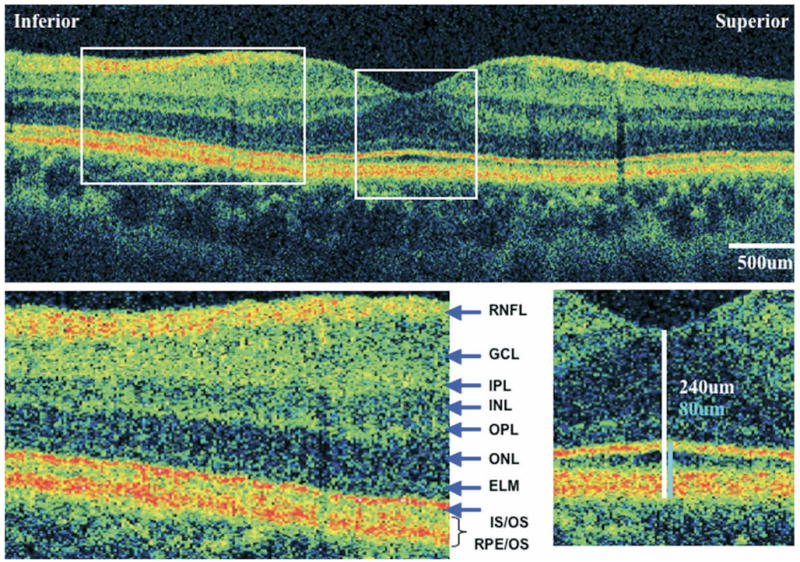
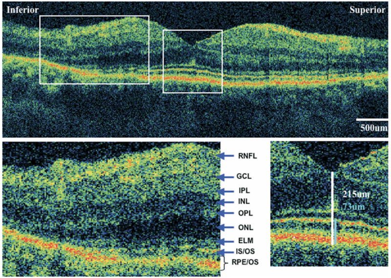
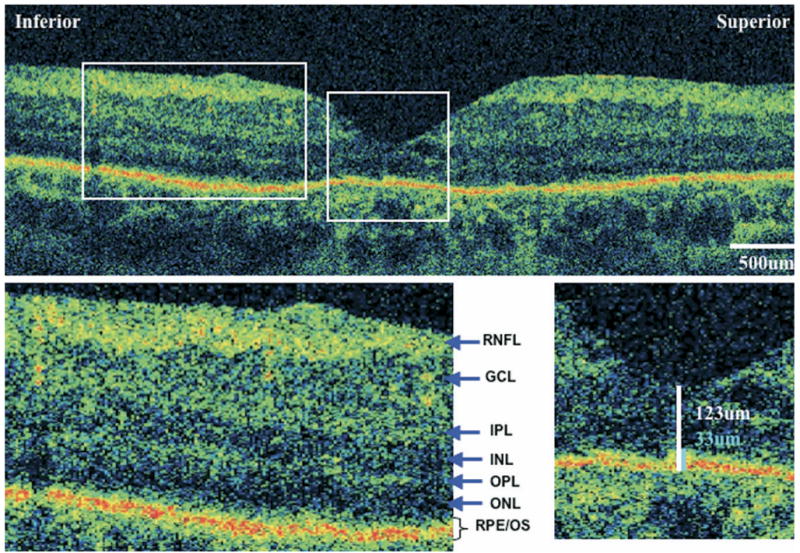
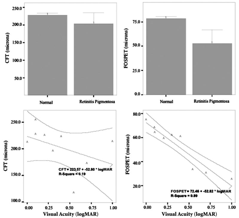
Purpose: To assess photoreceptor integrity in patients with retinitis pigmentosa (RP) and related diseases using ultra-high resolution optical coherence tomography (UHR-OCT) and to correlate foveal photoreceptor loss with visual acuity.
Design: Observational case series.
Methods: Nine eyes of nine patients with RP and related diseases were imaged with UHR-OCT at the ophthalmology clinic. Patients were diagnosed based on history, examination, fluorescein angiography, and electroretinography. Concurrently, 36 eyes of 36 normal subjects were imaged with UHR-OCT. Central foveal thickness (CFT) and foveal outer segment/pigment epithelium thickness (FOSPET) were defined and measured on UHR-OCT images in all subjects and were compared between the two groups using unpaired t tests. The two thickness measurements in RP patients were correlated with visual acuity using Pearson correlation and linear regression.
Results: UHR-OCT demonstrated macular photoreceptor thinning in all RP patients. The difference in CFT between RP patients and normal subjects was not statistically significant (P = .103), but the difference in FOSPET between the two groups was significant (P = .003). Visual acuity showed a fair correlation with CFT (Pearson r = -0.43, r(2) = 0.187, P = .245) and an excellent correlation with FOSPET (Pearson r = -0.942, r(2) = 0.887, P < .0001).
Conclusions: In the current study using UHR-OCT, a new thickness measurement termed FOSPET is demonstrated to quantify photoreceptor loss. FOSPET was statistically thinner in patients with RP and related diseases than in normal eyes and showed correlation with logMAR visual acuity. FOSPET appears to be a probable predictor of visual acuity in RP.
Figures
Similar articles
-
Assessment of central visual function in Stargardt's disease/fundus flavimaculatus with ultrahigh-resolution optical coherence tomography.Invest Ophthalmol Vis Sci. 2005 Jan;46(1):310-6. doi: 10.1167/iovs.04-0212. Invest Ophthalmol Vis Sci. 2005. PMID: 15623790
-
Autofluorescence imaging and spectral-domain optical coherence tomography in incomplete congenital stationary night blindness and comparison with retinitis pigmentosa.Am J Ophthalmol. 2012 Jan;153(1):143-54.e2. doi: 10.1016/j.ajo.2011.06.018. Epub 2011 Sep 13. Am J Ophthalmol. 2012. PMID: 21920492 Free PMC article.
-
Quantifying microstructural changes in retinitis pigmentosa using spectral domain - optical coherence tomography.Eye Vis (Lond). 2019 May 15;6:13. doi: 10.1186/s40662-019-0139-0. eCollection 2019. Eye Vis (Lond). 2019. PMID: 31123686 Free PMC article.
-
Vitamin A and fish oils for preventing the progression of retinitis pigmentosa.Cochrane Database Syst Rev. 2020 Jun 18;6(6):CD008428. doi: 10.1002/14651858.CD008428.pub3. Cochrane Database Syst Rev. 2020. PMID: 32573764 Free PMC article.
-
Diagnostic imaging in patients with retinitis pigmentosa.J Med Invest. 2012;59(1-2):1-11. doi: 10.2152/jmi.59.1. J Med Invest. 2012. PMID: 22449988 Review.
Cited by
-
Bilateral cystoid macular edema following docetaxel chemotherapy in a patient with retinitis pigmentosa: a case report.BMC Ophthalmol. 2015 Mar 29;15:32. doi: 10.1186/s12886-015-0020-4. BMC Ophthalmol. 2015. PMID: 25885440 Free PMC article.
-
Hyperautofluorescent ring in autoimmune retinopathy.Retina. 2012 Jul;32(7):1385-94. doi: 10.1097/IAE.0b013e3182398107. Retina. 2012. PMID: 22218149 Free PMC article.
-
Direct comparison of retinal structure and function in retinitis pigmentosa by co-registering microperimetry and optical coherence tomography.PLoS One. 2019 Dec 12;14(12):e0226097. doi: 10.1371/journal.pone.0226097. eCollection 2019. PLoS One. 2019. PMID: 31830067 Free PMC article.
-
Optical coherence tomography--current and future applications.Curr Opin Ophthalmol. 2013 May;24(3):213-21. doi: 10.1097/ICU.0b013e32835f8bf8. Curr Opin Ophthalmol. 2013. PMID: 23429598 Free PMC article. Review.
-
The findings of optical coherence tomography of retinal degeneration in relation to the morphological and electroretinographic features in RPE65-/- mice.PLoS One. 2019 Jan 29;14(1):e0210439. doi: 10.1371/journal.pone.0210439. eCollection 2019. PLoS One. 2019. PMID: 30695025 Free PMC article.
References
-
- Berson EL. Retinitis pigmentosa. The Friedenwald Lecture. Invest Ophthalmol Vis Sci. 1993;34:1659–1676. - PubMed
-
- Pagon RA. Retinitis pigmentosa. Surv Ophthalmol. 1988;33:137–177. - PubMed
-
- van Soest S, Westerveld A, de Jong PT, et al. Retinitis pigmentosa: defined from a molecular point of view. Surv Ophthalmol. 1999;43:321–334. - PubMed
-
- Milam AH, Li ZY, Fariss RN. Histopathology of the human retina in retinitis pigmentosa. Prog Retin Eye Res. 1998;17:175–205. - PubMed
-
- Milam AH, Li ZY, Cideciyan AV, Jacobson SG. Clinicopathologic effects of the Q64ter rhodopsin mutation in retinitis pigmentosa. Invest Ophthalmol Vis Sci. 1996;37:753–765. - PubMed
Publication types
MeSH terms
Grants and funding
LinkOut - more resources
Full Text Sources
Other Literature Sources
